Before reading the article below, read Part I:
Saving Smiles Beyond Implants: Minimally Invasive Solutions with Zirconia Cantilever Bridges – Part I

Fig.1
To address the challenge of “lost bonding” with zirconia, the gold standard surface treatment combines air abrasion with an MDP-based primer, enhancing both micromechanical interlocking and chemical bonding to resin. However, an alternative technique—glass-ceramic spray deposition—has also been shown to improve the bonding effectiveness of zirconia restorations.

Fig.2
This technique modifies the internal zirconia surface to achieve adhesion similar to that of glass ceramics. A lithium disilicate (LiDiSi) layer is mechanically bonded to the zirconia, enhancing its adhesion to dental cement.
Video.1
A lithium disilicate layer (Biomic LiSi Connect, Aidite) is sprayed onto the internal surface of pre-sintered zirconia (not in the green stage). During sintering, this process produces a thin, dense lithium disilicate layer (glass-melting effect) that penetrates the zirconia structure, creating a strong bond comparable to that of glass ceramics. Importantly, it does not alter the physical properties of zirconia during high-temperature crystallization, strengthening, or shrinkage, nor does it affect the final strength, material characteristics, or fit of the restoration.


Fig.3
Upon receiving the restorations, the provisional was removed and veneers were individually tried in to verify fit.

Fig.4
In the same way, veneers are collectively checked for interproximal contact and occlusal contact interferences.

Fig.5
In an occlusal view, restorations are assessed to ensure no palatal seating discrepancies.

Fig.6
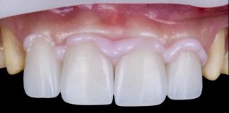
After verifying the fit and trying in the veneers, the different RBFDP designs were evaluated. The alternative design was tested first: although it provided a natural shade match, the connector’s position compromised the overall tooth aesthetics.

Fig.7
The restoration demonstrated natural blending and the optical effect of an individual tooth. The patient selected the no-prep RBFDP with a palatal wing design as the final treatment. Color was also evaluated.

Fig.8
For bonding, an adhesive protocol similar to that used for lithium disilicate was followed. After achieving absolute isolation with a rubber dam (NicTone Rubberdam Blue, medium thickness) and double autoligature, the restorations were cleaned with alcohol, etched with 9.5% hydrofluoric acid for 45–60 seconds, rinsed, dried, and treated with 37% phosphoric acid for 60 seconds to remove precipitates. Following a final rinse and drying, a ceramic primer (Monobond Plus) was applied.

Fig.9
After rubber dam and ligatures placement.

Fig.10
Dental tissues were air-abraded with 30-micron aluminum oxide.

Fig.11
Although in this case the RBFDP was cemented before the veneers, the preferred sequence is veneers first, then pontic. The palatal surface of tooth 21 was cleaned, sandblasted, etched with 37% phosphoric acid for 30 seconds, rinsed, and dried.

Fig.12
A primer was applied to the palatal enamel with a microbrush and air-dried, followed by a layer of unfilled adhesive (Heliobond), which was left uncured.

Fig.13
Variolink Esthetic LC Light was used for the RBFDP, which was seated with the aid of a 3D-printed positioning guide. The correct positioning of the guide and the RBFDP was verified both before and after rubber dam placement.

Fig.14
The guide should be designed over both the teeth and the planned restorations, providing support while allowing verification of correct fit and esthetic positioning. Ideally, a 3D-printed positioning guide should:
- Be rigid to ensure precise positioning.
- Be translucent to allow light-curing of the composite cement.
- Involve only the occlusal surfaces of adjacent teeth to avoid interference with the rubber dam, and include a squared window for clinical observation of the fit.
- Cover the buccal surface of the pontic to hold it securely in place.
- Provide one support on the wing and one on the buccal surface of the abutment tooth, while leaving all wing borders uncovered to facilitate removal of excess composite cement (the guide forms an arch at the incisal edge).

Fig.15
After removing the major excess cement, the RBFDP was spot-cured with a collimated light tip for 5–10 seconds to secure the restoration, after which the guide was removed.

Fig.16
The remaining teeth (12, 21, and 22) were etched with 37% orthophosphoric acid—15 seconds on dentin and 30 seconds on enamel. A dentin primer (Optibond FL Primer) was then applied, air-dried, and covered with an unfilled adhesive (Heliobond), which was left uncured.

Fig.17
Cement was applied internally, and the veneers were seated with finger pressure using Variolink Esthetic LC Light. Major excesses were removed from the interproximal, buccal, and palatal surfaces.

Fig.18
The veneers were spot-cured with a collimated light tip for 5 seconds from the palatal side. Interproximal excesses were then gently removed with dental floss, and an adhesive-coated brush was used to refine the buccal and palatal margins.

Fig.19
After removing excess material, an initial polymerization cycle of 20 seconds was performed. Final light-curing was then carried out under glycerin gel for 60 seconds per surface to prevent the formation of an oxygen-inhibited layer.

Fig.20
All the ligatures were removed, and margin polishing was performed with rubber points.

Fig.21
The rubber dam was removed, a fixed orthodontic retainer was placed, and the treatment outcome was evaluated.

Fig.22
After bonding, occlusal adjustments were performed with an Arkansas stone bur, followed by polishing with silicone rubbers specifically designed for zirconia. Occlusal contact with the pontic was not eliminated but slightly reduced, particularly in the distal area of the pontic and connector. Occlusal guidance was removed.

Fig.23
Buccal view after 6 weeks.

Fig.24
Lateral view after 6 weeks.

Fig.25
Esthetic result in full smile.

Fig.26
Esthetic result in rest position.

Fig.27
Conclusions
Zirconia veneers and cantilever RBFDPs represent excellent alternatives for achieving aesthetic integration and soft tissue stability. Their mechanical and optical properties make zirconia a promising material for minimally invasive rehabilitations, broadening treatment options beyond traditional ceramics.
To achieve a stable bond between zirconia and dental tissues, specific protocols such as the APC technique or internal surface modification with lithium silicate are essential to ensure long-term success. This case demonstrates how the use of digital workflows, connective tissue grafting, and advanced adhesive protocols led to predictable, aesthetic, and functionally stable results.
Bibliography
- Blatz MB, Alvarez C, Survye K, Brindis M. How to bond to zirconia: The APC Concept. J Adhes Dent. 2021;23(3):203-211. doi:10.3290/j.jad.a46003.
- Emmanouil-George C. Tzanakakis, DDS, MS, Ioannis G. Tzoutzas, DDS, MS, PhD, Petros T. Koidis, DDS, MS. Is there a potential for durable adhesion to zirconia restorations? A systematic review. J Prosthet Dent. 2016;115(1):9-19. doi:10.1016/j.prosdent.2015.06.007.
- Gerdolle D, Browet S, Gresnigt M. mPérennité des restaurations indirectes collées: « the no-finishing concept » Swiss Dent J. 2022 Jul 11;132(7-8):499-504. doi: 10.61872/sdj-2022-07-08-03.
- Gerdolle D, Browet S, Gresnigt M. Multi-luting concept: an adhesive protocol for indirect restorations to optimize bonding strength and longevity. Swiss Dent J. 2024 Feb 7;134(1):72-83. doi: 10.61872/sdj-2024-01-02.
- Gresnigt M, Jonker JA, van der Made SAM. The cantilever contact-point resin bonded bridge; adhesion 2.0. J Esthet Restor Dent. 2024;36(1):37-46. doi:10.1111/jerd.13179.
- Tezulas E, Yildiz C, Evren B, Ozkan Y. Clinical procedures, designs, and survival rates of all-ceramic resin-bonded fixed dental prostheses in the anterior region: A systematic review. J Esthet Restor Dent. 2018;30:307–318. systematic review. J Esthet Restor Dent. 2018;00:1–12. https://doi.org/10.1111/jerd.12389.
- Jonker JA, Tirlet G, Dagba A, Marniquet S, Ouwerkerk M , Cune MS, Gresnigt M.A 32-month evaluation of lithium disilicate cantilever resin-bonded fixed dental prostheses to replace a missing maxillary incisor. J Prosthet Dent 2024;132:956-963.
- Gurel G. Predictable, precise, and repeatable tooth preparation for porcelain laminate veneers. Pract Proced Aesthet Dent. 2003;15(1):17-24; quiz 26.
- Gurel G. The Science and Art of Porcelain Laminate Veneers. Chicago: Quintessence Publishing Co; 2003.
- Kern M, Gläser R. Single-retainer all-ceramic resin-bonded fixed dental prostheses: Long-term outcomes in the esthetic zone. J Esthet Restor Dent. 2023;35(1):64-73. doi:10.1111/jerd.13001.
- Kern M, Türp L, Yazigi C. Long-term outcome of anterior cantilever zirconia ceramic resin-bonded fixed dental prostheses: Influence of the pontic location. J Prosthet Dent. 2025;133(8):1017-1023.
- Mainjot AKJ. No-Prep Zirconia Cantilever Resin-Bonded Fixed Dental Prostheses: A Noninvasive, Simple Approach to Replacing a Single Missing Tooth. J Esthet Restor Dent. 2025;37(1):68-84.
- Magne P, Belser UC. Novel porcelain laminate preparation approach driven by a diagnostic mock-up. J Esthet Restor Dent. 2004;16(1):7-16; discussion 17-18. doi:10.1111/j.1708-8240.2004.tb00002.x.
- Mekled S, Elwazeer S, Jurado CA, White J, Faddoul F, Afrashtehfar KI, Fischer NG. Ultra-Translucent Zirconia Laminate Veneers: The Influence of Restoration Thickness and Stump Tooth-Shade. J Esthet Restor Dent. 2022;34(2):279-291. doi:10.1111/jerd.12801.
- Tzanakakis EG, Tzoutzas IG, Koidis PT. Is there a potential for durable adhesion to zirconia restorations? A systematic review. J Prosthet Dent. 2016;115(1):9-19. doi:10.1016/j.prosdent.2015.06.007.
- Kang, C.-M.; Lin, D.-J.; Feng, S.-W.; Hung, C.-Y.; Iwaguro, S.; Peng, T.-Y. Innovation Glass-Ceramic Spray Deposition Technology Improving the Adhesive Performance for Zirconium-Based Dental Restorations. Int. J. Mol. Sci. 2022,23,12783. https://doi.org/ 10.3390/ijms232112783.
- Peng TY, Kang MC, Feng SW, Hung CY, Iwaguro S, Lin DJ. Effects of glass-ceramic spray deposition manipulation on the surface characteristics of zirconia dental restorations. Ceramics International, https://doi.org/10.1016/j.ceramint.2022.06.252.