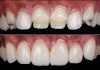
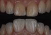
Currently, the dental community is highly focused on minimally invasive restorative procedures. In cases of tooth agenesis or tooth loss due to trauma, caries, or periodontal disease, treatment options include removable prostheses, orthodontic space closure, cantilever resin-bonded fixed dental prostheses (RBFDP), conventional bridges, and implant-supported crowns. Implant therapy is often the first choice in adult patients. However, implant failures still occur, and although reintervention is possible, treatment decisions must prioritize the patient’s perspective, who often exhibit reluctance due to prior negative experiences. Managing scenarios where patients do not consent to implant regeneration and placement is among the most challenging in aesthetic dentistry, thus treatment must be guided by minimally invasive procedures and prosthetic options.
The most prominent representatives of this minimally invasive approach are veneers and cantilever RBFDPs. The most commonly used materials for these restorations are lithium disilicate and feldspathic ceramics. Zirconia is increasingly gaining attention as a promising material for such treatments. Nonetheless, many clinicians remain skeptical, relegating zirconia primarily to implant-supported prostheses or cases demanding high mechanical retention, such as crowns, due to concerns that resin cement adhesion might fail. Despite being non-etchable, protocols like the APC concept: (A) airparticle abrasion, (P) zirconia primer, and (C) adhesive composite resin; or internal surface modification with lithium silicate (LiSi) have demonstrated stable and durable adhesion. Zirconia offers an excellent alternative for both veneers and cantilever RBFDPs, combining high aesthetics, minimal thicknesses, and the option of high translucency. Mechanically and physically, zirconia presents advantages over other ceramics: failures tend to be adhesive rather than cohesive fractures, giving a second chance to rebond the material to the tooth and it provides an effective solution to the challenge of blending materials to achieve optimal aesthetic results—an obstacle in all aesthetic rehabilitations. Several factors influence material selection, including restoration type (implant-supported, tooth-supported, or hybrid), stump color, retention mode (traditional crown, biologically oriented preparation technique [BOPT] crown, veneer, onlay, or RBFDPs), and tissue type (enamel, dentin, cementum), among others. With zirconia, once protocols for reliable adhesion are established, many of these issues are resolved, enabling more aesthetic and simpler, albeit not simplistic treatments.


Fig.1
In this article, we present the case of a 25-year-old patient with agenesis of the maxillary right central incisor (tooth 11), who came to our clinic following regenerative and implant failure. She had worn a removable partial denture throughout adolescence. At the age of 24, orthodontic treatment was carried out, followed by GBR and immediate implant placement, which became infected and failed. A provisional PMMA Maryland bridge was then provided.

Fig.2
Facial and intraoral photographs were taken to assess the aesthetics and the soft tissue defect.

Fig.3
CBCT was performed to assess the extent of the defect at the bone level, observing the bone peaks.

Fig.4
Due to her previous negative experience, the patient declined further implant surgery. A minimally invasive aesthetic approach was therefore proposed, combining a connective tissue graft with veneers and a cantilever RBFDP. Before initiating the definitive treatment, the patient’s smile aesthetics were improved by correcting the incisal canting caused by the provisional RBFDP, which was recontoured using Sof-Lex discs.

Fig.5
The palatal wing was reduced to involve only tooth 21.

Fig.6
For the connective tissue graft, two timing modalities exist: a prior approach, performed before bonding, or an immediate approach, conducted concurrently with RBFDP bonding either the same day or within the following week. The immediate approach is currently preferred because it promotes soft tissue healing influenced by prosthesis design. However, in this case, due to a severe defect, the graft was performed before the definitive prosthesis. The temporary Maryland was removed.

Fig.7
A connective tissue graft was harvested from the palate.

Fig.8
The graft was de-epithelialized with a 15C blade to obtain pure connective tissue. A partial flap was then elevated, and the graft was stabilized in a saddle-like position with sutures to ensure stability and optimal healing.

Fig.9
The graft healed in place for two months without pressure. During this period, the prosthetic tooth was used as a temporary pontic, stabilized with an Essix retainer.

Fig.10
Controlled pressure was then applied to the soft tissue by modifying the removable pontic with flowable and packable composite to create an ovate pontic. This pressure induced mild gingival ischemia, causing brief discomfort but no pain, which resolved within five minutes—indicating that the pressure was moderate and insufficient to cause soft tissue necrosis.

Fig.11
A one-week interval was maintained between adjustments, during which the soft tissue was gradually displaced interproximally, buccally, and apically.

Fig.12
When the gingival margins of the pontic 11 and tooth 21 were aligned, pressure application ceased. This marked a total of four weeks of moderate gingival modification, followed by one month of soft tissue stabilization.

Fig.13
Intraoral scanning was performed during the stabilization phase, and a digital wax-up was created by the dental technician using Exocad software. Two designs were prepared, the first including a pontic at tooth 11 and a veneer for tooth 21.

Fig.14
The second design included a pontic at tooth 11 and veneers on teeth 12, 21, and 22. The final treatment plan consisted of ceramic veneers on teeth 12, 21, and 22, combined with a cantilever RBFDP at tooth 11.

Fig.15
The iTero Element scanner was used to capture the maxillary arch with the mock-up in place, along with the mandibular arch and the bite registration.

Fig.16
The Additive Preparation Technique (APT), as described by Dr. Galip Gurel, was employed. Preparation was performed through the mock-up using calibrated burs to create guided grooves, ensuring adequate veneer thickness.

Fig.17
The grooves were marked with a graphite pencil before removing the mock-up. The marked areas were then reduced until the marks disappeared, and the surface was homogenized using a straight green-coded bur.

Fig.18
After completing the incisal and vestibular preparation, the cervical margin was refined with a small round bur. The orthodontist was asked to leave the necessary space on the palatal aspect of tooth 21. Although several cantilever RBFDP designs exist, preparation was avoided in favor of more conservative approaches, namely the No-Prep Cantilever RBFDP and the Cantilever Contact-Point RBFDP, the latter showing promising fracture resistance comparable to full crowns.

Fig.19
Zirconia from Aidite was selected for both veneers and pontics. Although lithium disilicate and feldspathic ceramics remain the gold standard for veneers due to their well-documented adhesion, they generally require minimal thicknesses of 0.2 mm cervical to 1.5 mm incisal. For RBFDPs, lithium disilicate demands connector dimensions of 3 mm in width and 4 mm in height, with wing thicknesses between 1–1.2 mm. Failures in these materials typically present as ceramic chipping, fractures, or wing debonding. Zirconia has emerged as a strong alternative, offering superior mechanical and aesthetic properties while requiring reduced dimensions: veneer thicknesses of 0.2–0.4 mm and RBFDPs with 0.5 mm wing retainers and connectors of 2 mm width and 3 mm height. With a reported 15-year survival rate of 97%, zirconia allows for more conservative and less invasive treatments.

Fig.20
To ensure uniform and minimal restoration thickness, an incisal preparation control silicone guide was used.

Fig.21
A sagittal preparation control silicone guide was also used.

Fig.22
After verifying thickness and insertion axis, photographs were taken with and without polarizers using VITA shade guide tabs and Natural Die Color tabs to document stump shade and achieve accurate color matching.

Fig.23
Polytetrafluoroethylene (PTFE) tape was used as a retraction cord in the sulcus. The preparations and cervical margins were then refined with red-coded burs and polished using Enhance points and Sof-Lex discs.

Fig.24
Preparations were scanned with the PTFE in place. Teflon tape offers advantages as a retraction material due to its hydrophobic properties and lack of shape memory, allowing it to adapt closely to the sulcus and facilitate margin visualization without interfering during scanning.

Fig.25
Margins were immediately marked, and all data were sent to the dental technician. Immediate post-scan margin marking is a key advantage of digital workflows, enabling intraoral verification and reducing the risk of errors.

Fig.26
A PMMA temporary was fabricated using the “egg shell” technique.

Fig.27
The PMMA restoration was intraorally sandblasted, treated with a PMMA adhesive (Anaxblend, Anaxdent), and relined with Structur bisacryl resin using a 3D-printed positioning guide. After relining, the excess material was removed and the provisional was polished.

Fig.28
The zirconia selected in collaboration with the laboratory for both veneers and pontics was Aidite EZneer High Value Multilayered, a 5Y-TZP ultra-high translucency zirconia ideal for minimal-thickness veneers.

Fig.29
Prosthetic restorations fabricated by Rafael Jaén TPD.

Fig.30
For the pontic at tooth 11, an alternative design was also milled. In cases combining a pontic with veneers, the “Laminate Veneer Retainer” (LVR) design uses the adjacent veneer as the pontic wing. Both designs—the no-prep RBFDP with a palatal veneer and the RBFDP with an LVR—are shown. As observed, the LVR appears less natural because the connector’s palatal positioning remains visible on the buccal side. For this reason, the LVR design is generally limited to lateral incisor pontics, where the connector can be hidden behind the distal contour of the central incisor.

Fig.31
To accurately replicate the desired color, the technician applied the Natural Die technique, fabricating a composite stump to match the target shade and fine-tune the integration between the underlying tooth, adjacent dentition, and final restoration. It is important to note, for some zirconia brands, the final shade for laminate veneers with varying thicknesses is significantly influenced by the stump shade, but often in ways contrary to common assumptions. For example, the thinnest zirconia veneers (0.5 mm) tend to appear lighter (B1 shade) over backgrounds ranging from A1 to A4, while thicker veneers (0.75 mm and 1.0 mm) tend to display darker shades (B2). These findings have crucial implications for achieving optimal aesthetics with zirconia veneers of varying thicknesses.
Continue reading in Part II:
Saving Smiles Beyond Implants: Minimally Invasive Solutions with Zirconia Cantilever Bridges – Part II
Bibliography
- Blatz MB, Alvarez C, Survye K, Brindis M. How to bond to zirconia: The APC Concept. J Adhes Dent. 2021;23(3):203-211. doi:10.3290/j.jad.a46003.
- Emmanouil-George C. Tzanakakis, DDS, MS, Ioannis G. Tzoutzas, DDS, MS, PhD, Petros T. Koidis, DDS, MS. Is there a potential for durable adhesion to zirconia restorations? A systematic review. J Prosthet Dent. 2016;115(1):9-19. doi:10.1016/j.prosdent.2015.06.007.
- Gerdolle D, Browet S, Gresnigt M. mPérennité des restaurations indirectes collées: « the no-finishing concept » Swiss Dent J. 2022 Jul 11;132(7-8):499-504. doi: 10.61872/sdj-2022-07-08-03.
- Gerdolle D, Browet S, Gresnigt M. Multi-luting concept: an adhesive protocol for indirect restorations to optimize bonding strength and longevity. Swiss Dent J. 2024 Feb 7;134(1):72-83. doi: 10.61872/sdj-2024-01-02.
- Gresnigt M, Jonker JA, van der Made SAM. The cantilever contact-point resin bonded bridge; adhesion 2.0. J Esthet Restor Dent. 2024;36(1):37-46. doi:10.1111/jerd.13179.
- Tezulas E, Yildiz C, Evren B, Ozkan Y. Clinical procedures, designs, and survival rates of all-ceramic resin-bonded fixed dental prostheses in the anterior region: A systematic review. J Esthet Restor Dent. 2018;30:307–318. systematic review. J Esthet Restor Dent. 2018;00:1–12. https://doi.org/10.1111/jerd.12389.
- Jonker JA, Tirlet G, Dagba A, Marniquet S, Ouwerkerk M , Cune MS, Gresnigt M.A 32-month evaluation of lithium disilicate cantilever resin-bonded fixed dental prostheses to replace a missing maxillary incisor. J Prosthet Dent 2024;132:956-963.
- Gurel G. Predictable, precise, and repeatable tooth preparation for porcelain laminate veneers. Pract Proced Aesthet Dent. 2003;15(1):17-24; quiz 26.
- Gurel G. The Science and Art of Porcelain Laminate Veneers. Chicago: Quintessence Publishing Co; 2003.
- Kern M, Gläser R. Single-retainer all-ceramic resin-bonded fixed dental prostheses: Long-term outcomes in the esthetic zone. J Esthet Restor Dent. 2023;35(1):64-73. doi:10.1111/jerd.13001.
- Kern M, Türp L, Yazigi C. Long-term outcome of anterior cantilever zirconia ceramic resin-bonded fixed dental prostheses: Influence of the pontic location. J Prosthet Dent. 2025;133(8):1017-1023.
- Mainjot AKJ. No-Prep Zirconia Cantilever Resin-Bonded Fixed Dental Prostheses: A Noninvasive, Simple Approach to Replacing a Single Missing Tooth. J Esthet Restor Dent. 2025;37(1):68-84.
- Magne P, Belser UC. Novel porcelain laminate preparation approach driven by a diagnostic mock-up. J Esthet Restor Dent. 2004;16(1):7-16; discussion 17-18. doi:10.1111/j.1708-8240.2004.tb00002.x.
- Mekled S, Elwazeer S, Jurado CA, White J, Faddoul F, Afrashtehfar KI, Fischer NG. Ultra-Translucent Zirconia Laminate Veneers: The Influence of Restoration Thickness and Stump Tooth-Shade. J Esthet Restor Dent. 2022;34(2):279-291. doi:10.1111/jerd.12801.
- Tzanakakis EG, Tzoutzas IG, Koidis PT. Is there a potential for durable adhesion to zirconia restorations? A systematic review. J Prosthet Dent. 2016;115(1):9-19. doi:10.1016/j.prosdent.2015.06.007.
- Kang, C.-M.; Lin, D.-J.; Feng, S.-W.; Hung, C.-Y.; Iwaguro, S.; Peng, T.-Y. Innovation Glass-Ceramic Spray Deposition Technology Improving the Adhesive Performance for Zirconium-Based Dental Restorations. Int. J. Mol. Sci. 2022,23,12783. https://doi.org/ 10.3390/ijms232112783.
- Peng TY, Kang MC, Feng SW, Hung CY, Iwaguro S, Lin DJ. Effects of glass-ceramic spray deposition manipulation on the surface characteristics of zirconia dental restorations. Ceramics International, https://doi.org/10.1016/j.ceramint.2022.06.252.