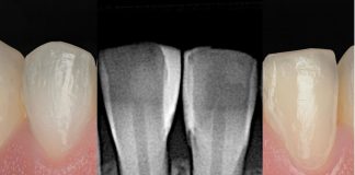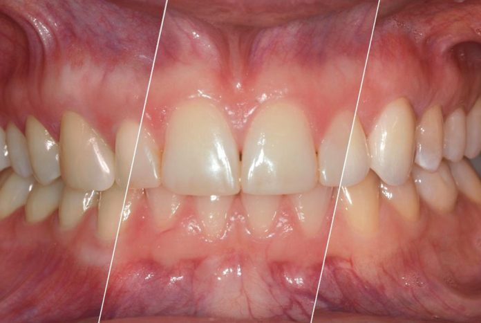
A clinical case by our Community member Dr. Alexander Strunz
This article and its content are published under the Author’s responsibility as an expression of the Author’s own ideas and practice. Styleitaliano denies any responsibility about the visual and written content of this work.
Aesthetic rehabilitation of the anterior dentition presents a particular challenge, especially in young patients. In addition to functional considerations, atient expectations regarding esthetics, as well as the integration of previous treatments, play a crucial role in treatment planning.
The following case presents the interdisciplinary management of a young female patient who presented to our clinic after completing orthodontic treatment, seeking aesthetic improvement of unaesthetic anterior composite restorations. The patient’s history of previous treatment for temporomandibular disorder (TMD) further emphasized the need to carefully balance functional stability with esthetic outcomes.
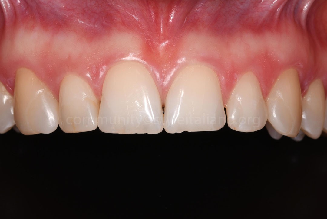
Fig.1
At the initial consultation, the patient presented following completion of orthodontic treatment. The existing anterior composite restorations exhibited noticeable color mismatch, and the patient was particularly dissatisfied with the shape and overall appearance of her anterior teeth. Although she initially expressed a preference for an indirect restorative approach, a combined direct–indirect treatment concept was selected.
Given the anticipated changes to the dynamic occlusal relationships and the patient’s history of successfully treated temporomandibular disorder (TMD), this minimally invasive staged approach was considered the most appropriate. The objective was to evaluate and, if necessary, refine the functional implications of the planned morphological modifications over time before transferring the definitive tooth shape to ceramic veneers.
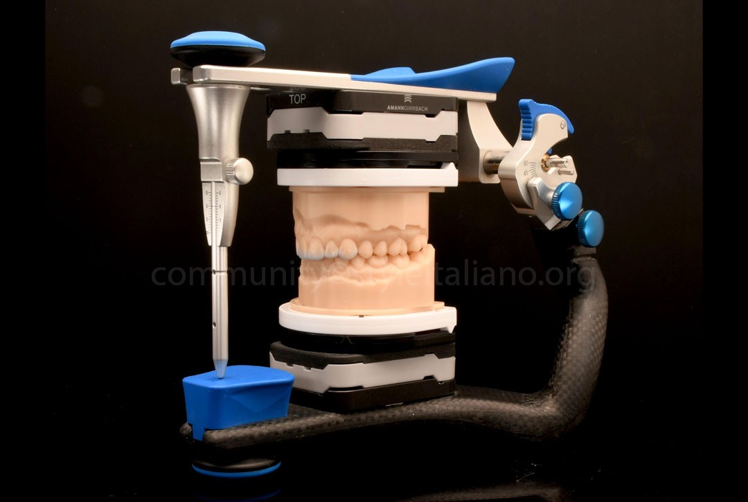
Fig.2
Following an intraoral scan of the initial clinical situation, study models were fabricated using 3D printing and subsequently mounted in an articulator.
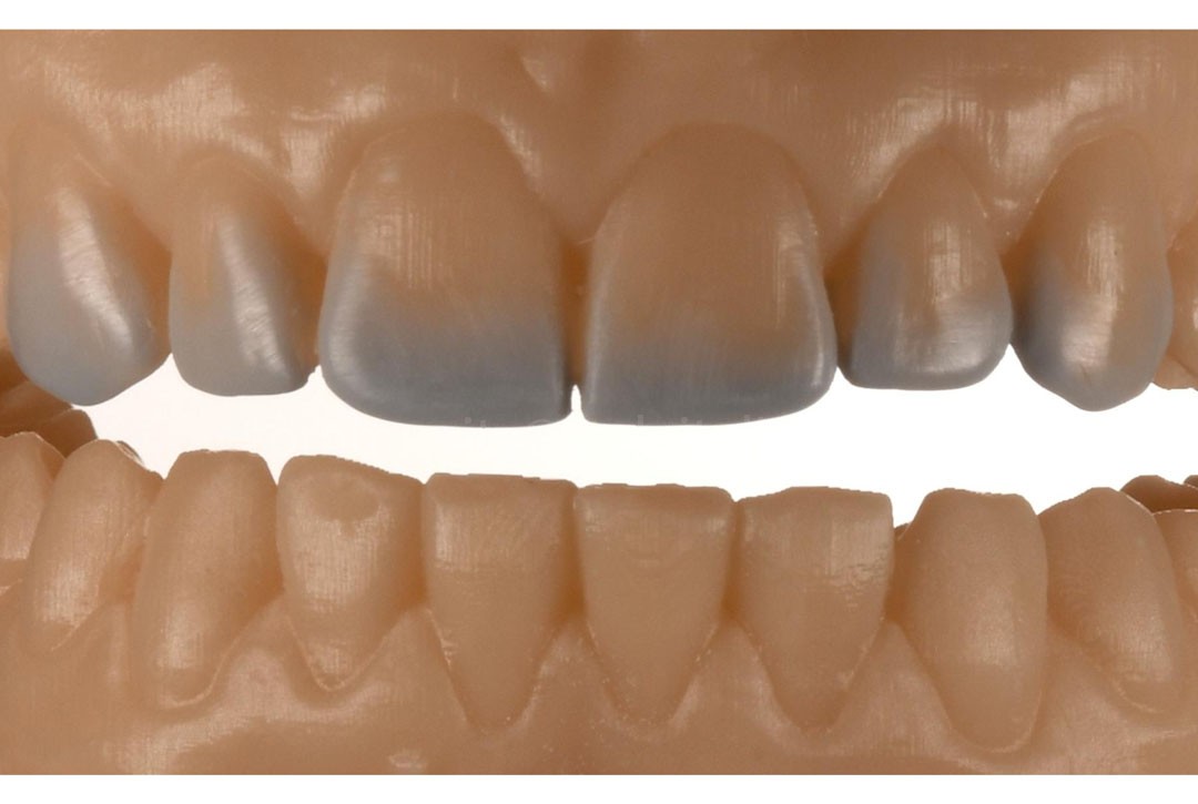
Fig.3
The diagnostic wax-up was then completed by the clinician on these models. This step was intentionally performed using a conventional analog workflow to provide the clinician with a tactile and visual understanding of the planned morphological modifications before proceeding with the restorative treatment.
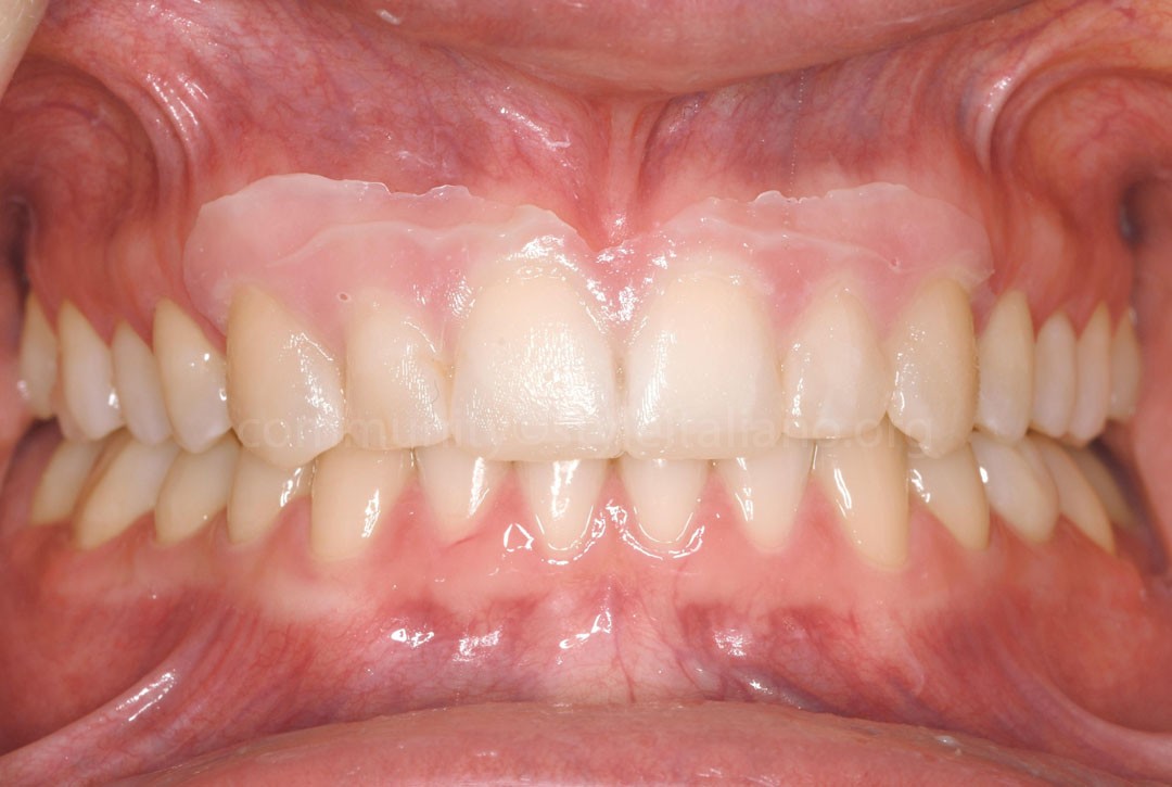
Fig.4
Using a silicone index, the proposed morphological changes were transferred intraorally as a mock-up with provisional resin, allowing the patient to visualize and evaluate the proposed treatment outcome. Once the patient was satisfied with the reshaping, the provisional composite material was removed. Using a silicone index, the morphology of the palatal wall was recorded.
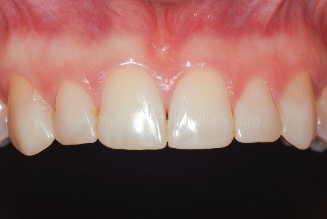
Fig.5
After the patient approved the proposed design, the existing anterior composite restorations were removed. The planned modifications in tooth shape were then implemented using a direct composite approach, establishing the intended esthetic and functional contours as an intermediate stage before the definitive ceramic restorations.
Figure 5 illustrates the clinical appearance of the direct composite restorations at the 6-month follow-up. No complications related to the modified occlusal scheme were observed during the follow-up period. Minor occlusal adjustments were performed in the regions of teeth 13 and 23 at the 3-, 6-, and 12-month recall appointments to further optimize functional guidance and patient comfort.
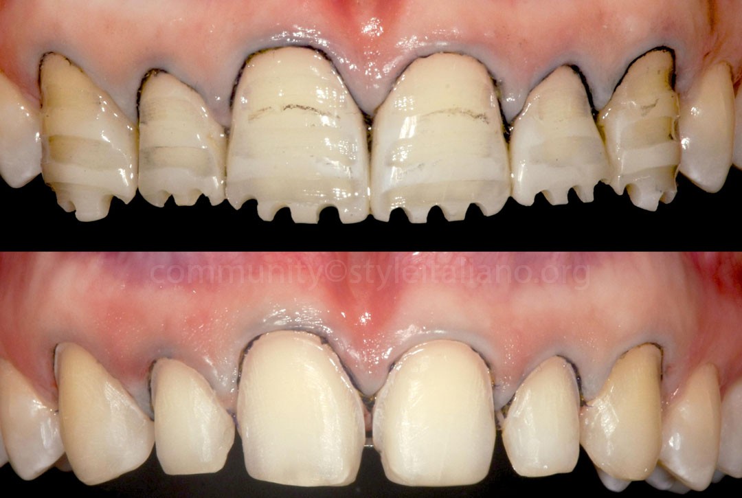
Fig.6
After a preoperative digital scan (pre-scan), preparation for the definitive ceramic veneers was initiated approximately one year after the start of treatment. Depth-cutting burs were used to achieve a uniform incisal reduction of 1.0 mm and a circumferential reduction of 0.5 mm, ensuring adequate space for the ceramic restorations while preserving tooth structure.
Placement of retraction cords during tooth preparation facilitated visualization and precise positioning of the preparation margins while respecting the principles of the supracrestal tissue attachment.
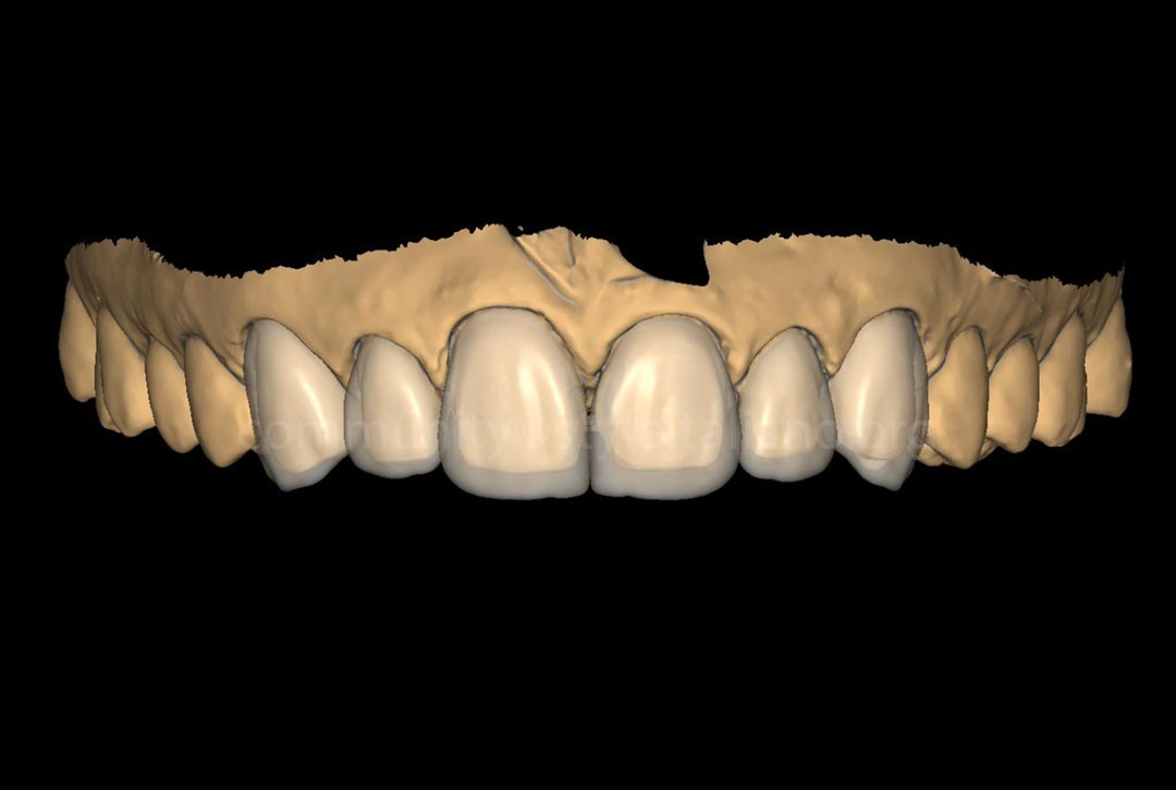
Fig.7
A second intraoral scan was obtained after tooth preparation with the retraction cords in place, the definitive restorations were fabricated using a digital laboratory workflow (Schwandner H).
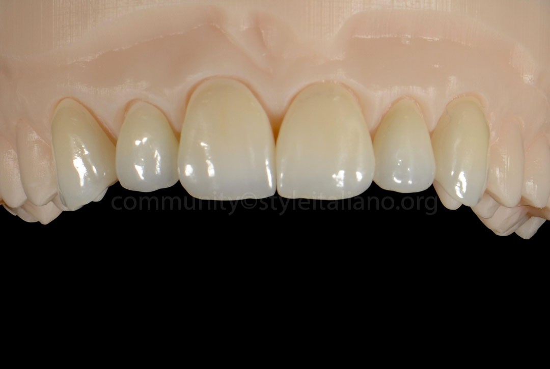
Fig.8
Monolithic zirconia veneers were designed and milled using CAD/CAM technology, followed by individual characterization through staining and glazing to achieve the desired esthetic outcome.
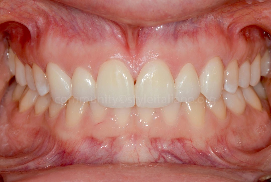
Fig.9
Immediate postoperative view following final cementation.
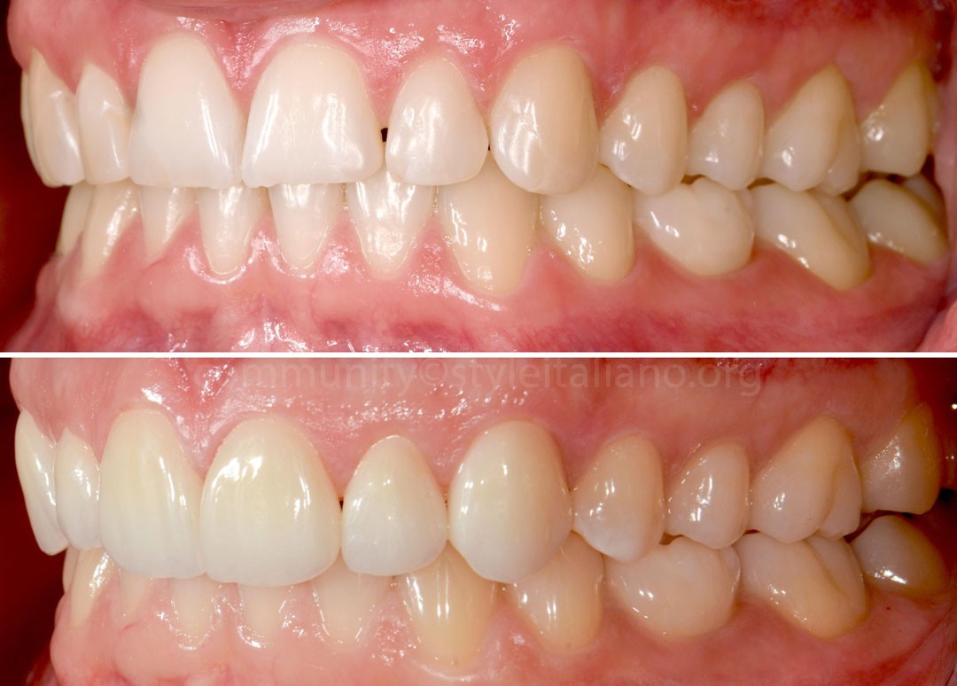
Fig.10
Conclusions
This case demonstrates that a staged direct–indirect treatment concept can provide a predictable, minimally invasive approach for the esthetic rehabilitation of the anterior dentition in patients with complex functional histories. Interim direct composite restorations allowed the planned morphological changes to be evaluated and refined under functional conditions before definitive ceramic rehabilitation. Combined with a digital workflow and monolithic zirconia veneers, this approach resulted in a predictable esthetic and functional outcome while preserving tooth structure.
Bibliography
- Fradeani M, Barducci G. Esthetic Rehabilitation in Fixed Prosthodontics. Vol. 2: Prosthetic Treatment: A Systematic Approach to Esthetic, Biologic and Functional Integration. Quintessence Publishing; 2008.
- Putignano A, Cerutti A, Mangani F. Guidelines for Adhesive Dentistry: The Key to Success. Quintessence Publishing; 2009.
- Schwartz-Arad D. Esthetics in Dentistry. Quintessence Publishing; 2016.