A clinical case by our Community member Dr. Tuan Anh Nghiem
This article and its content are published under the Author’s responsibility as an expression of the Author’s own ideas and practice. Styleitaliano denies any responsibility about the visual and written content of this work.
Restoring anterior teeth affected by enamel hypoplasia requires balancing high aesthetic expectations with the preservation of healthy tooth structure. One of the main clinical challenges is achieving predictable adhesion, as hypomineralized areas often present islands of sound enamel. Conventional preparation strategies may therefore result in unnecessary removal of healthy tissue.
To address this challenge, a digitally driven workflow based on Digital Smile Design (DSD) was implemented to predict and visualize the desired aesthetic outcome. Based on this digital blueprint, limited orthodontic movement was performed to optimize tooth position and reduce the need for restorative preparation, while periodontal plastic surgery was used to refine the gingival architecture and enhance pink aesthetics. Finally, a mock-up guided preparation protocol allowed for controlled reduction while ensuring seamless aesthetic and functional integration.
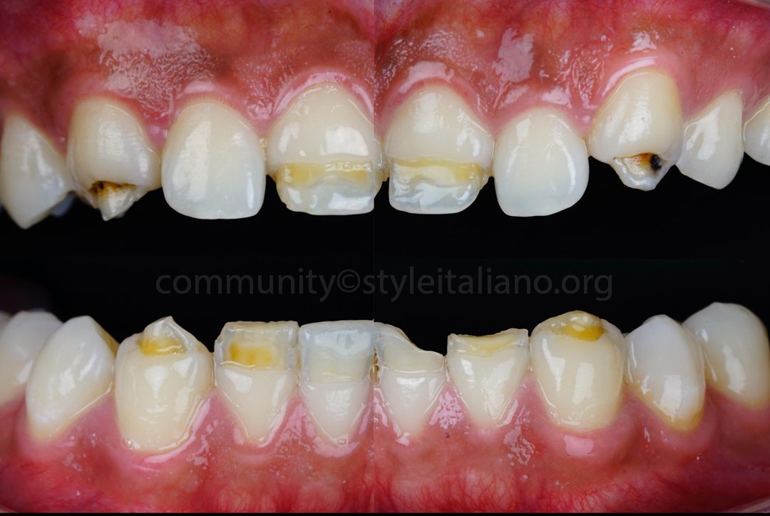
Fig.1
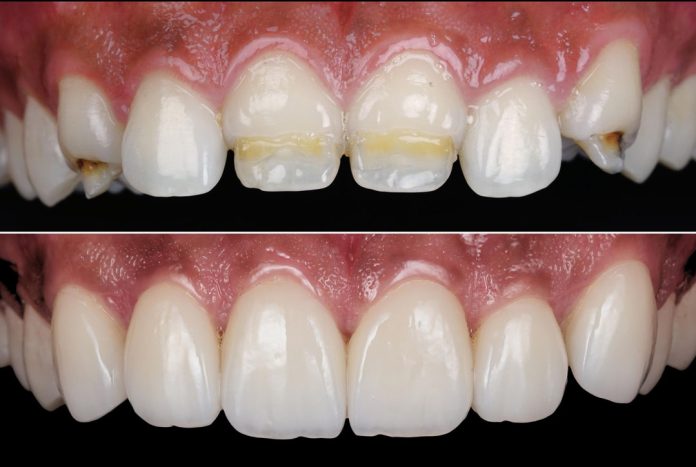
A young male patient presented with significant aesthetic concerns related to extensive enamel hypoplasia affecting the anterior dentition. Clinical examination revealed active carious lesions on the hypoplastic structures of 13 and 23. Additionally, structural loss was observed on 31 and 32. The complex combination of enamel defects, decay, and fractures severely affected both the aesthetics and structural integrity of the anterior zone.
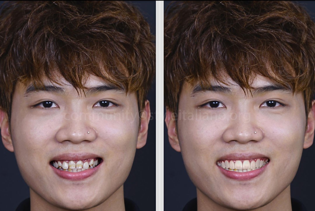
Fig.2
To perform a comprehensive facial analysis, we evaluated his widest smile display. Digital Smile Design (DSD) was utilized to simulate the future smile.
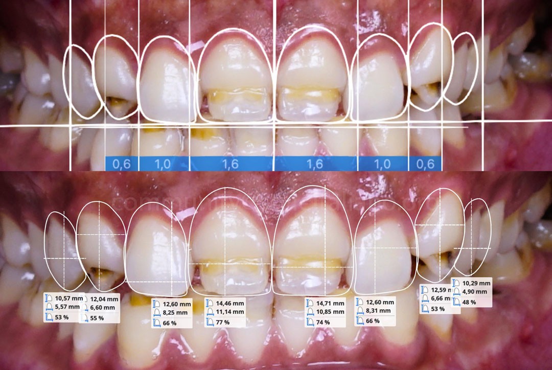
Fig.3
Digital Smile Design (DSD) was utilized to establish a precise reference for the ideal tooth morphology and gingival contours, with proportions tailored to the patient’s face and expectations. This digital blueprint served as a diagnostic compass, allowing the patient to preview the aesthetic outcome while guiding the upcoming orthodontic and periodontal treatment phases.
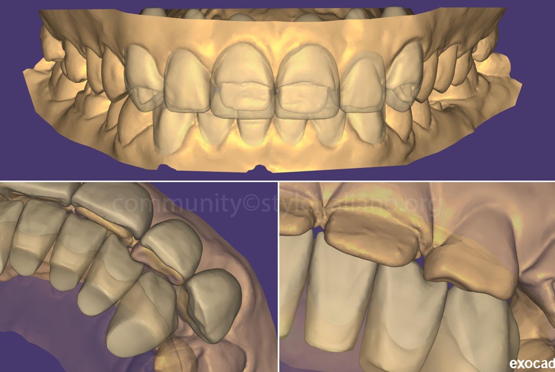
Fig.4
Based on the patient’s intraoral scans, a digital wax-up was designed to simulate the final tooth morphology, position, and the required preparation depth. Analyzing this digital wax-up revealed a critical clinical insight: without pre-restorative treatment, significant tooth reduction would be unavoidable, especially on tooth 22. The uncorrected positioning of the anterior teeth was highly unfavorable for conservative adhesive restorations, confirming the absolute need for prior intervention.
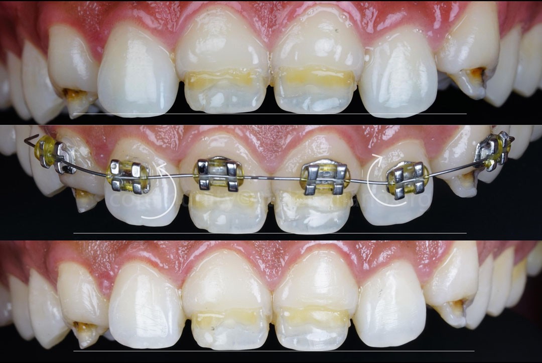
Fig.5
To overcome this challenge, limited orthodontics was initiated to intrude teeth 12 and 22, as well as to correct their axial rotation. This targeted tooth movement aimed at realiging them into an ideal position within the arch. By repositioning these teeth slightly, we could optimize the restorative space and establish a favorable path of insertion, dramatically reducing the need for aggressive tooth preparation.
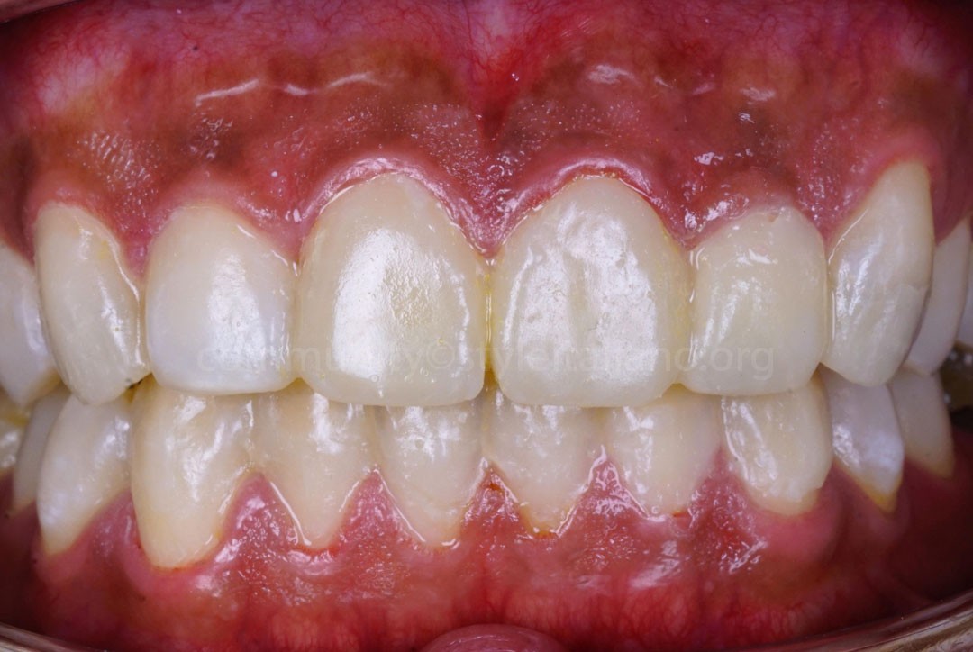
Fig.6
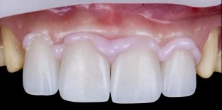
Following the limited orthodontic phase, the patient’s smile aesthetics were re-evaluated by transferring the digital wax-up data into a direct intraoral mock-up. Combining this clinical mock-up with the initial DSD analysis, it became evident that the crown height remained sub-optimal. To achieve ideal tooth proportions and balance, periodontal plastic surgery for clinical crown lengthening was required before proceeding to the final restoration.
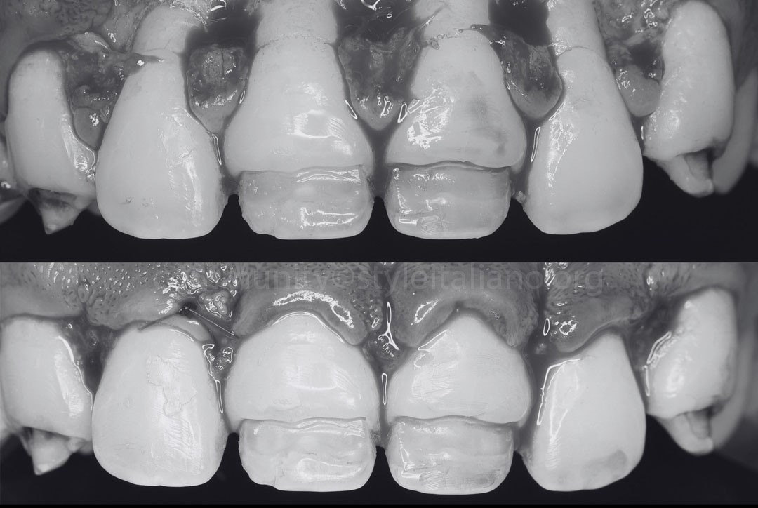
Fig.7
To ensure long-term stability and prevent gingival relapse, a conventional gingivectomy was avoided. Instead, an apically positioned flap (APF) combined with minor osseous recontouring was performed in the maxillary anterior region. This approach effectively elongated the clinical crowns while maintaining a healthy biologic width. The surgical site was meticulously sutured, and a 6-week healing period was allowed for soft tissue maturation.
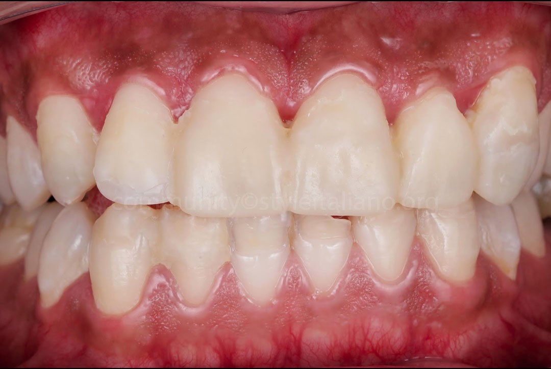
Fig.8
To actively guide and condition the healing gingival architecture, a temporary mock-up was delivered to the maxillary anterior region. This early provisionalization acted as a physical matrix, driving the tissue to heal precisely around the planned contours. The margins of the mock-up were meticulously finished and highly polished to minimize bacterial adhesion, preventing plaque accumulation and ensuring optimal soft tissue health during the healing phase.
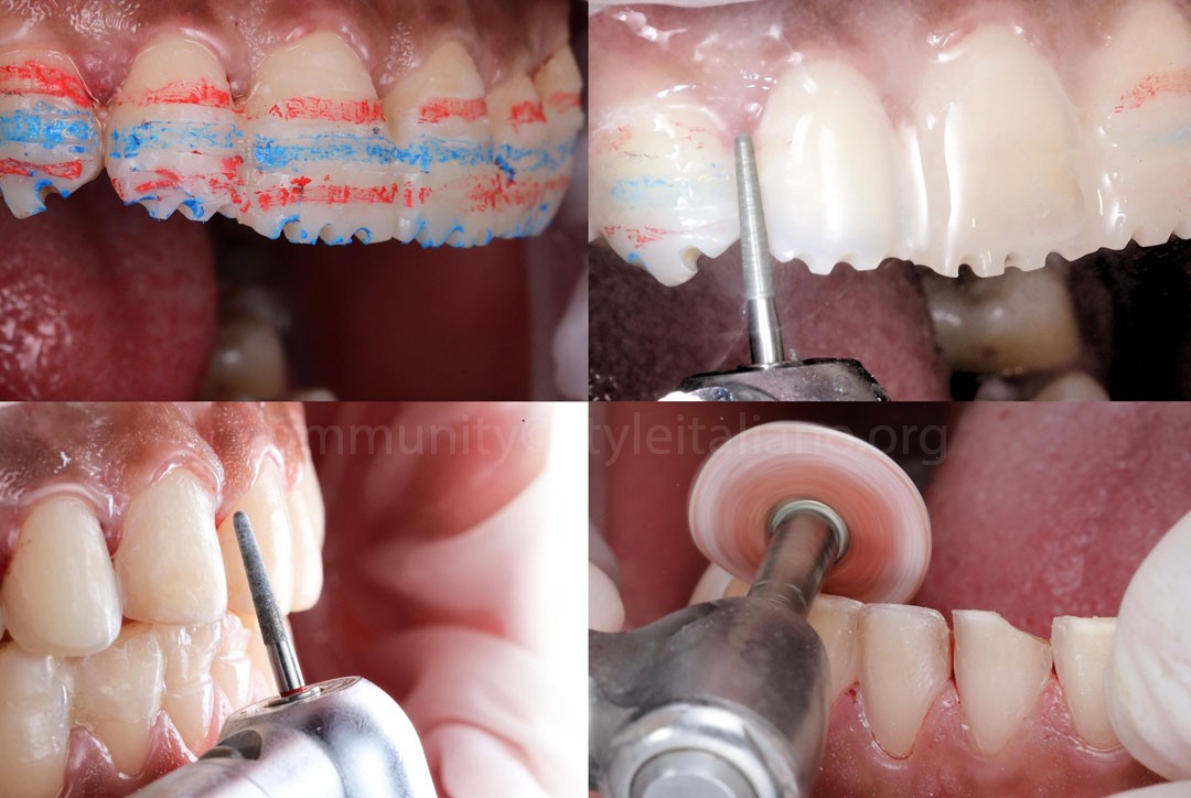
Fig.9
Following the healing period, a minimally invasive mock-up guided preparation protocol was executed. Depth-orientation burs were used to create precise reduction guides of 0.5mm on the labial surface and 1mm at the incisal edge. Concurrently, active carious tissues were selectively removed using a round bur, ensuring maximum preservation of the adjacent sound tooth structure. Once the minimally invasive preparation was completed, the margins and surfaces were pre-polished using a coarse Sof-Lex disc to eliminate sharp angles.
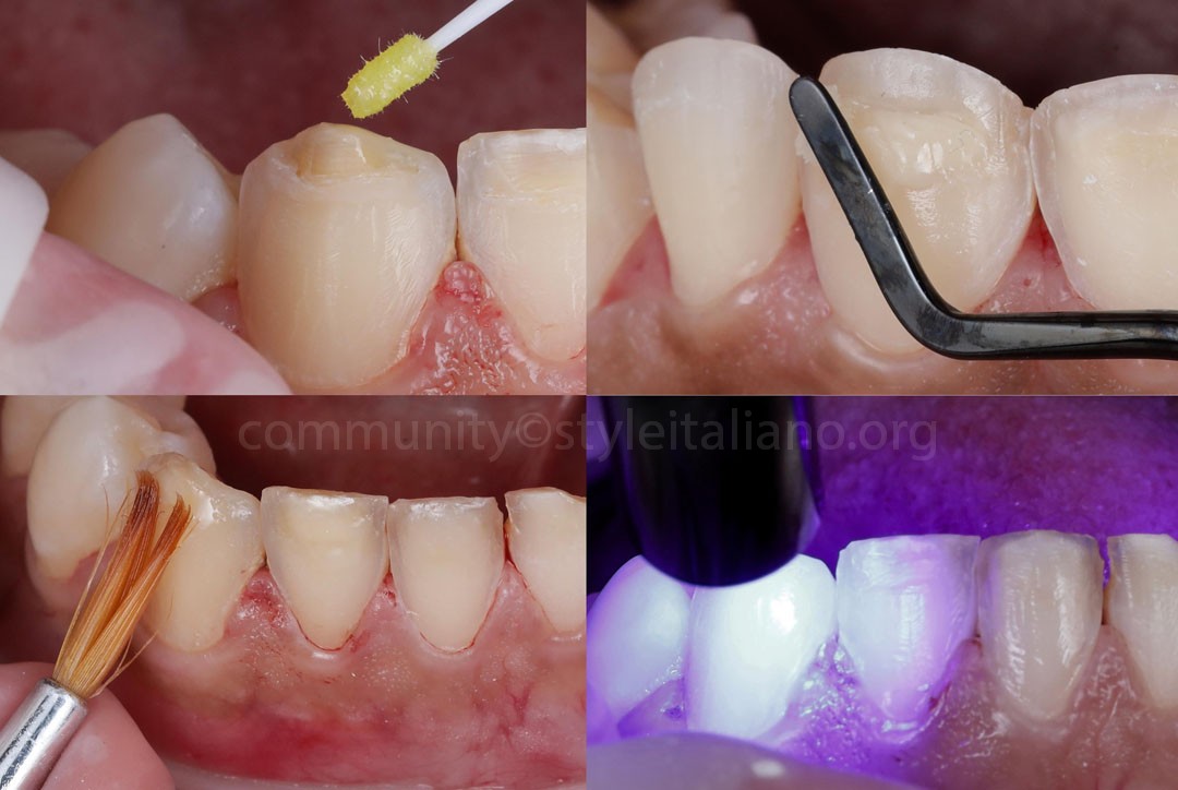
Fig.10
In areas where dentin was exposed following caries removal within the hypoplastic substrate, an Immediate Dentin Sealing (IDS) protocol was performed. Subsequently, an opaque composite layer was placed to restore surface continuity and mask the underlying discolored dentin. This step not only maximized bond strength and protected the pulp but also established a neutralized optical background for the final aesthetic restoration.
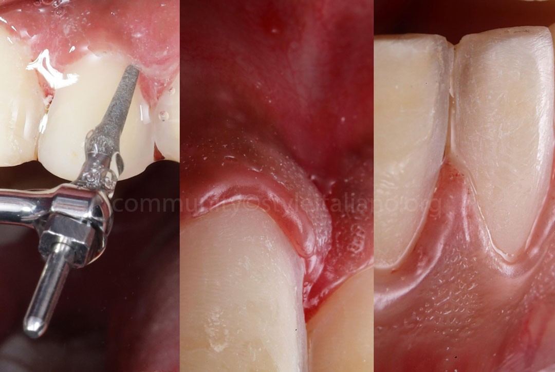
Fig.11
To finalize the preparations, ultrasonic tips were utilized for margin refinement and surface smoothing. This ultrasonic finishing ensured highly defined, sharp preparation lines while eliminating any micro-irregularities and avoiding soft tissue trauma. With the margins perfectly sharpened and prepped, the substrate was completely optimized for a high-precision impression.
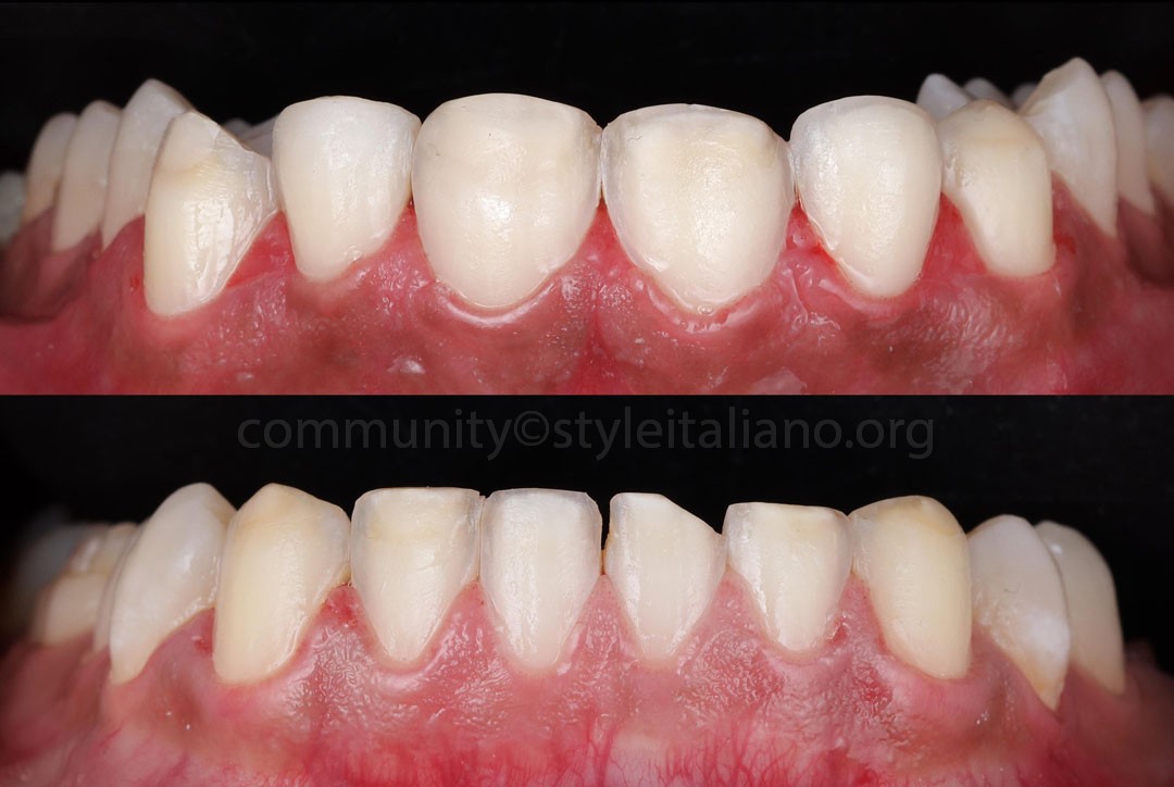
Fig.12
The ultra-conservative preparations demonstrate highly defined, ultrasonically refined margins and smooth surface topography.
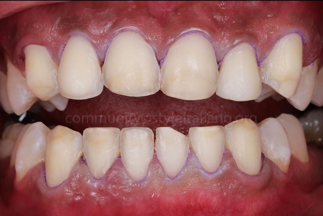
Fig.13
To ensure maximum accuracy for the final impression, gingival retraction cords were carefully placed in the sulcus of all prepared teeth. This step provided temporary displacement of the free gingiva, completely exposing the ultrasonically refined margins and allowing for an unobstructed capture of the finish lines.
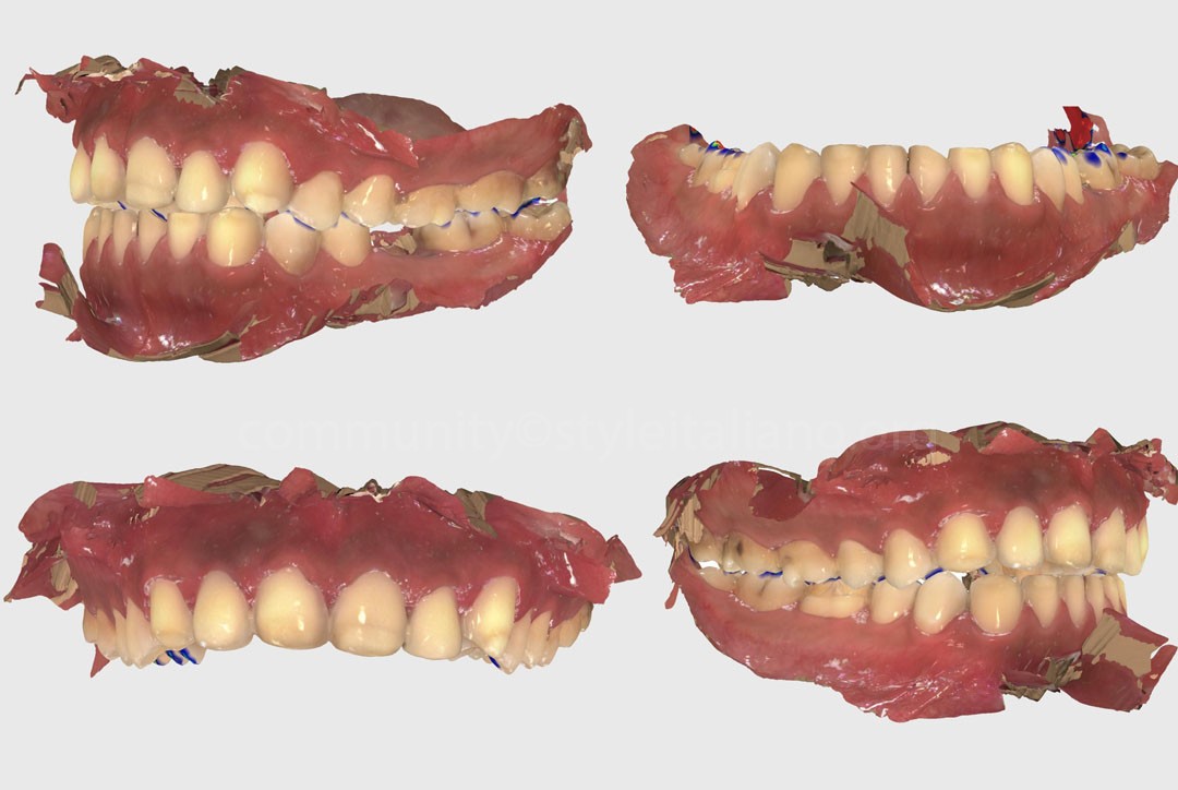
Fig.14
High-precision digital impressions of both arches were then captured using the Primescan intraoral scanner (Dentsply Sirona).
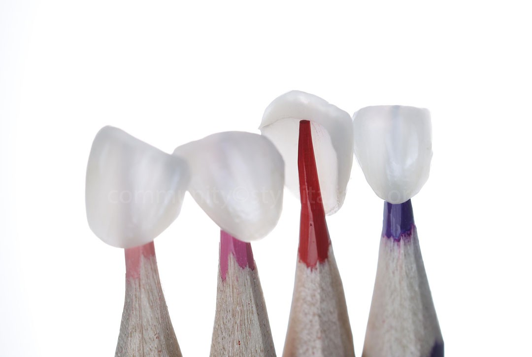
Fig.15
Lithium disilicate glass-ceramic veneers (Initial LiSi, GC) were fabricated to restore the anterior teeth. This material was selected for its exceptional optical properties, high-definition surface details, and lifelike biomimetic aesthetics.
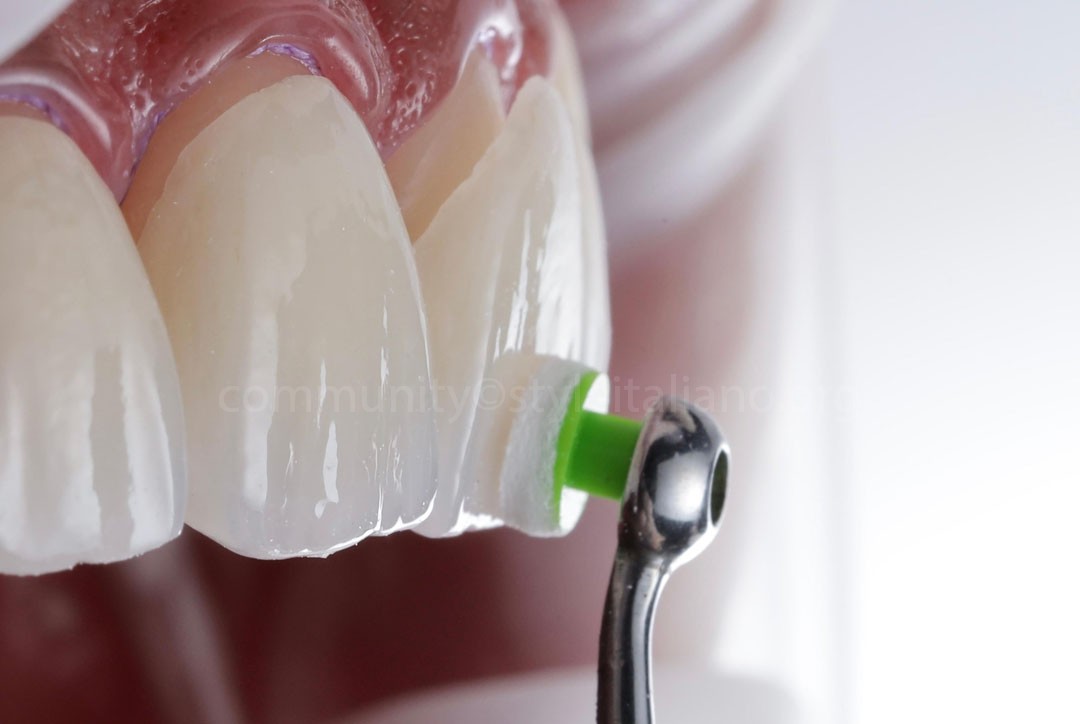
Fig.16
During the try-in phase, the glass-ceramic veneers exhibited excellent optical integration with the surrounding dentition.
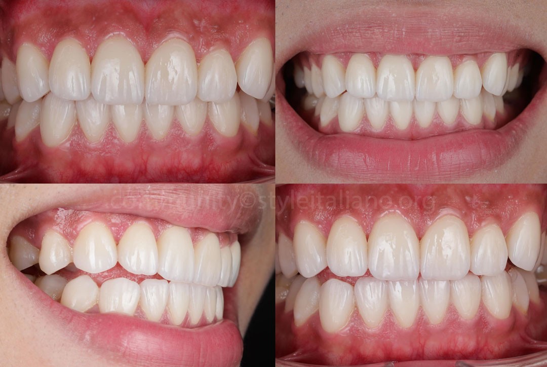
Fig.17
Post-operative view immediately after final cementation.
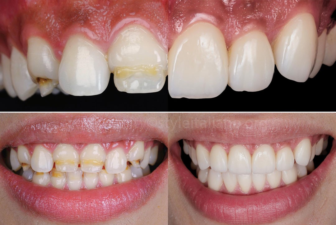
Fig.18
Before and after comparison showcasing the natural aesthetic transition.
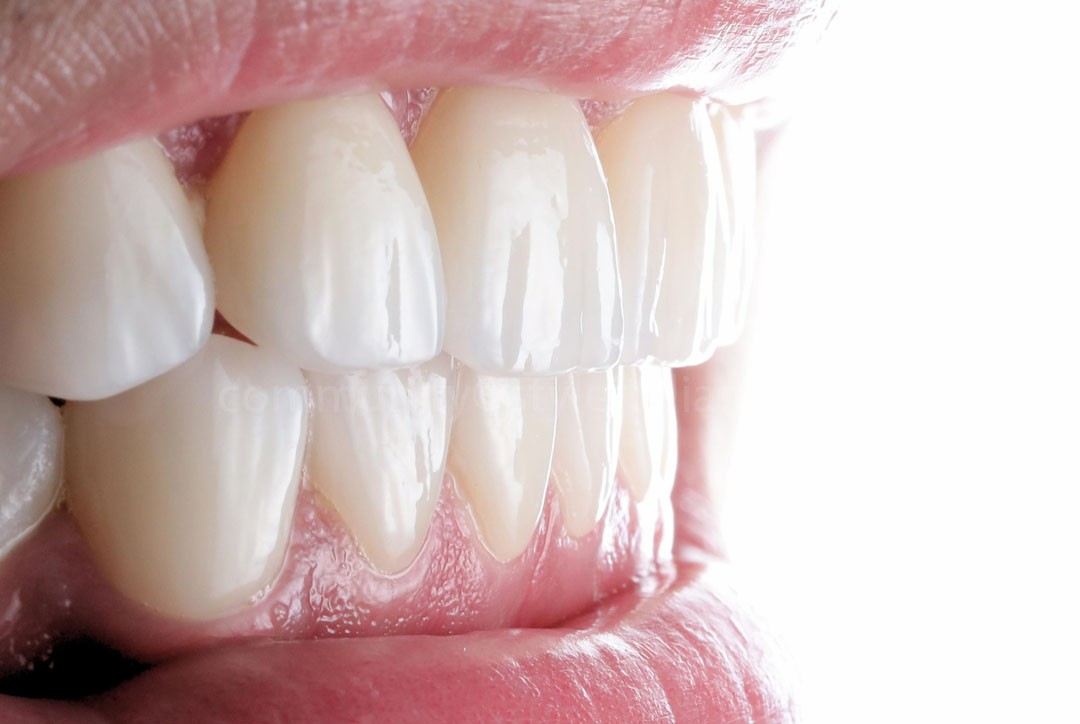
Fig.19
Close-up view highlighting the detailed surface optical effects.
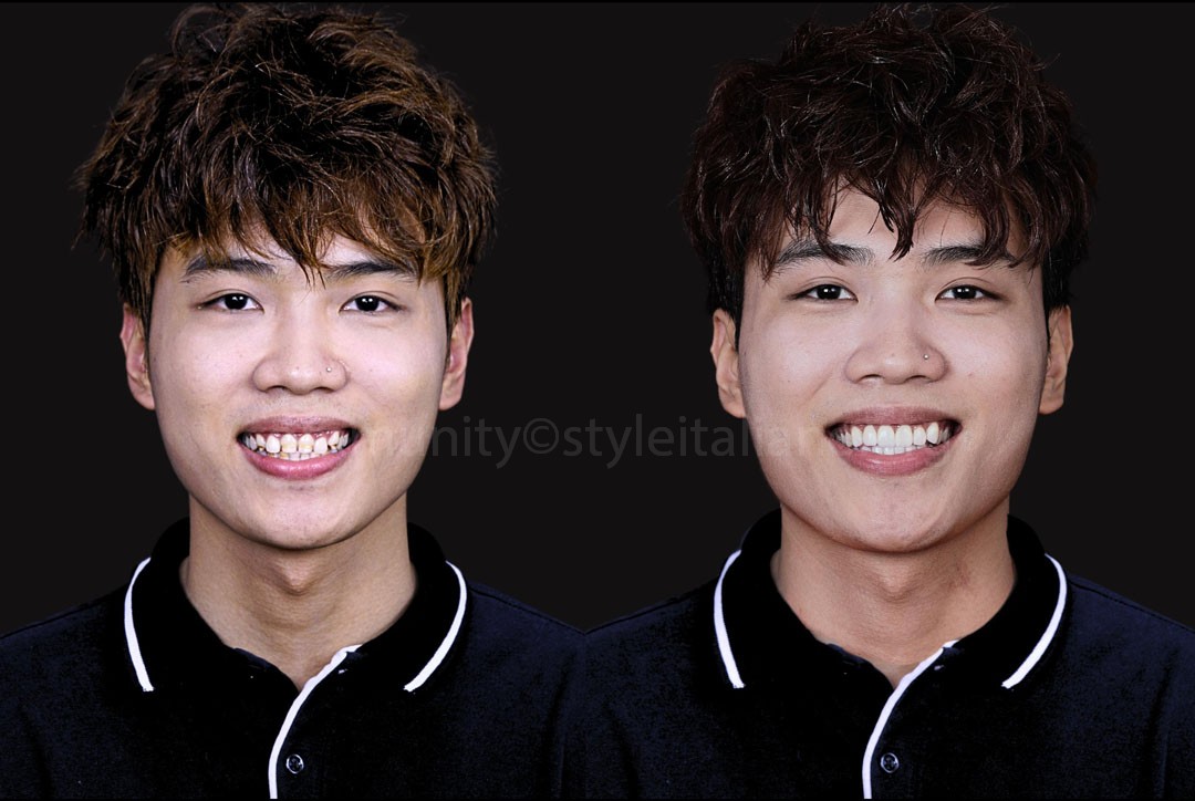
Fig.20
Extra-oral full smile views showcasing the dramatic aesthetic improvement.
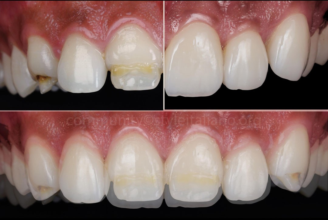
Fig.21
Conclusions
Achieving predictable and natural-looking outcomes in patients affected by structural enamel defects often requires treatment strategies that extend beyond restorative procedures alone. This case highlights the value of a digitally driven interdisciplinary workflow in which Digital Smile Design (DSD), limited orthodontic treatment, and periodontal plastic surgery were integrated to optimize tooth position and gingival architecture before restorative intervention.
The combination of mock-up guided minimally invasive preparation, Immediate Dentin Sealing (IDS), and lithium disilicate restorations enabled the resolution of both aesthetic and functional challenges while preserving the maximum amount of healthy tooth structure. A systematic and tissue-preserving approach such as this can improve treatment predictability and support long-term biomimetic outcomes.
Bibliography
- Magne P, Belser U. Bonded Porcelain Restorations in the Anterior Dentition: A Biomimetic Approach. Quintessence Publishing; 2002.
- Lasserre F, Magne M. FUSION: Art and Nature in Ceramic Restorations; Fundamentals / Clinic and Laboratory. Quintessence Publishing; 2014.
- Gürel G, Morimoto S, Calamita MA, Coachman C, Sesma N. Clinical performance of porcelain laminate veneers: outcomes of the aesthetic pre-evaluative temporary (APT) technique. International Journal of Periodontics & Restorative Dentistry. 2012;32(6):625-635.
- Vianna AL, Prado M, Dias KR, de Castro AM. Conservative aesthetic management of severe enamel hypoplasia: A multidisciplinary approach. Journal of Esthetic and Restorative Dentistry. 2017;29(3):157-164.
- Coachman C, Calamita MA. Digital Smile Design: A Tool for Treatment Planning and Communication in Esthetic Dentistry. Quintessence Dental Technology (QDT). 2012;35:103-111.