Indirect bonding with self-adhesive restorative materials can be challenging, particularly when multiple restorations are being bonded within the same arch, where efficient workflow and ease of cleanup are critical. One of the main clinical difficulties is determining the optimal timing for removing excess cement. Premature removal may risk dislodging the restoration, whereas delayed removal can result in hardened cement remnants, especially in interproximal areas, making cleanup difficult and time-consuming.
This article presents a challenging full-mouth reconstruction restored with zirconia crowns and describes a detailed clinical workflow, with particular emphasis on the bonding procedure.


Fig.1
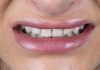
A 52-year-old female patient presented with a chief complaint of persistent gingival bleeding, halitosis, and soreness of the masticatory muscles. She was also dissatisfied with the shape of her existing crowns and their monochromatic appearance.
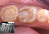
Clinical examination revealed that she had undergone full-mouth rehabilitation three years prior; however, multiple restorations exhibited significant deficiencies. In particular, inadequate marginal adaptation and poorly sealed finish lines were observed, contributing to gingival inflammation and spontaneous bleeding.

Fig.2
Clinical examination revealed a deep bite associated with a loss of vertical dimension, resulting in a collapsed occlusion.

Fig.3
In the first phase of treatment, periodontal health was addressed across the dentition. A soft tissue laser was used for decontamination of the gingival pockets. Additionally, restoration margins were carefully refined and polished with a diamond bur to approximate the finish lines as closely as possible. These procedures facilitated initial healing of the periodontal tissues.

Fig.4
Provisional PMMA crowns were fabricated for the maxillary incisors, as these teeth were the most affected. Retaining the existing crowns would have hindered optimal soft tissue healing; therefore, their replacement with well-contoured temporaries allowed for improved periodontal recovery.

Fig.5
The provisional crowns were cemented using Temp-Bond™ Clear (Kerr Corporation, Orange, California, USA) and maintained in situ for a period of four months to allow for adequate soft tissue healing and stabilization.

Fig.6
During the healing phase, a definitive treatment plan was formulated based on the newly established vertical dimension and centric relation. A bite registration was obtained after three weeks of deprogramming with a Kois deprogrammer, allowing accurate recording of the maxillomandibular relationship.

Fig.7
A new occlusal scheme was digitally designed using exocad, in accordance with the restored vertical dimension and the newly established centric relation record.

Fig.8
After four months with the provisional crowns, the periodontal tissues demonstrated healthy conditions, with no signs of inflammation and no bleeding on probing.

Fig.9
The definitive maxillary and mandibular crowns were fabricated using zirconia. Facial layering was applied from the first premolar to the contralateral first premolar to enhance esthetic integration in the visible zone, while the second premolars and molars were restored with monolithic zirconia to optimize strength and functional performance.

Fig.10
The definitive maxillary and mandibular crowns were fabricated using zirconia, with facial layering limited to the esthetic zone extending from the first premolar to the contralateral first premolar to enhance optical properties and esthetic integration.

Fig.11
Following the try-in procedure with liquid glycerin, the zirconia crowns were thoroughly rinsed with water and gently air-abraded using 27 μm aluminum oxide particles. The air pressure was maintained below 3 bar to avoid excessive surface alteration. This cleaning protocol aimed to remove potential salivary phosphate contamination and optimize the zirconia surface for adhesive cementation.

Fig.12
Following air abrasion, the zirconia crowns were thoroughly cleaned with water and medical-grade alcohol to remove residual contaminants and prepare the surfaces for subsequent adhesive cementation procedures.

Fig.13
After cleaning, the zirconia crowns were thoroughly dried, and a universal zirconia conditioning primer containing the 10-methacryloyloxydecyl dihydrogen phosphate (10-MDP) functional monomer was applied. The primer was left undisturbed on the internal surface of the crowns for 2 minutes, followed by gentle air drying to promote effective chemical bonding with the zirconia substrate.

Fig.14
On the day of definitive bonding, the periodontal tissues demonstrated complete maturation and optimal health, with no signs of inflammation or bleeding. The teeth were cleaned with water followed by chlorhexidine application. The exposed enamel margins were selectively etched with phosphoric acid for 30 seconds to enhance enamel bonding prior to adhesive cementation.

Fig.15
The definitive zirconia crowns were cemented using Maxcem Elite™ Chroma (Kerr Corporation, Orange, California, USA), a dual-cure, self-etching, self-adhesive resin cement. This cement provides a visual indication of the setting process through a color change, facilitating the identification of the optimal time for excess cement removal during the bonding procedure.

Fig.16
Through a reduction–oxidation reaction, the pink color of the Maxcem Elite™ Chroma gel point indicator gradually fades as polymerization progresses due to its patented redox initiator system. In addition to providing reliable adhesion, color stability, and fluoride release, this cement offers a valuable clinical advantage by providing a visual indication of the optimal timing for excess cement removal.

Fig.17
When Maxcem Elite™ Chroma transitions from a light pink color to an off-white appearance, the optimal gel phase for excess cement removal is reached. Excess cement was easily removed using instruments such as Excesso (LM-Dental, Parainen, Finland) and interproximal finishing metal strips. The color-change technology provides a reliable visual indication of the appropriate working time, allowing a simple, efficient, and predictable cleanup procedure. The excess cement can be removed with minimal effort, leaving clean restoration margins with minimal residual material.

Fig.18
Following excess cement removal, a clear glycerin gel was applied to the labial and palatal margins of the restorations to inhibit the oxygen-inhibited layer. Additional light polymerization was then performed for 40 seconds on each surface using a curing light to ensure complete polymerization of the marginal resin cement.

Fig.19
Two weeks after insertion, minor occlusal adjustments were performed on several occasions to achieve optimal functional harmony. Clinical evaluation demonstrated excellent soft tissue healing, with healthy periodontal tissues and a favorable biological response to the newly placed crowns.

Fig.20
Two weeks after insertion, the occlusion remained stable at the newly established vertical dimension and centric relation. The tissues demonstrated an excellent biological response to the definitive prosthesis, with healthy and well-adapted soft tissue conditions.

Fig.21
The new occlusal scheme resulted in improved muscular relaxation and a more harmonious facial appearance, with enhanced proportions of the lower third of the face following restoration of the vertical dimension.

Fig.22
Two weeks after definitive prosthesis insertion, the patient presented with a confident smile and excellent periodontal health. A clear thermoplastic night guard was provided for routine use to protect the hard and soft tissues and minimize functional stresses on the newly restored dentition.

Fig.23
Two weeks after definitive prosthesis insertion, the patient demonstrated a confident smile with excellent esthetic integration of the restorations and healthy periodontal tissues. A clear pressed thermoplastic night guard was provided for routine use to protect the hard and soft tissues and minimize functional stresses on the restored dentition.

Fig.24
Two weeks after definitive prosthesis insertion, the patient exhibited a harmonious smile that complemented her facial characteristics and personality. The restorations were designed and positioned to follow the natural smile line, achieving an esthetic integration between the dentition, facial features, and overall appearance.
Conclusions
The use of a color-changing self-etching resin cement, combined with appropriate adhesive protocols, facilitates predictable and efficient excess cement removal. This feature is particularly advantageous when bonding multiple restorations simultaneously, as it provides the clinician with a clear indication of the optimal cleanup time.
This simplified and controlled cementation procedure contributes to well-integrated restorations with excellent biological compatibility, reduced risk of postoperative sensitivity, and enhanced potential for long-term clinical success.
Bibliography
- Tosco V, Monterubbianesi R, Orilisi G, Sabbatini S, Conti C, Özcan M, Putignano A, Orsini G. Comparison of two curing protocols during adhesive cementation: can the step luting technique supersede the traditional one? Odontology. 2021 Apr;109(2):433-439. doi: 10.1007/s10266-020-00558-0. Epub 2020 Oct 31. PMID: 33128650; PMCID: PMC7954706.
- Koubi S, Raskin A, Dejou J, About I, Tassery H, Camps J, Proust JP. Effect of dual cure composite as dentin substitute on the marginal integrity of Class II open-sandwich restorations. Oper Dent. 2010 Mar-Apr;35(2):165-71.
- Blatz MB, Sadan A, Kern M. Resin-ceramic bonding: a review of literature. J Prosthet Dent 2003;89:268-74.