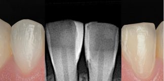
In managing deep carious lesions where dentin excavation approaches the pulp—especially in teeth with significantly compromised structure—an indirect pulp capping protocol should be carefully considered. In these scenarios, preserving the pulpal vitality and restoring the structural integrity of the tooth become equally critical.
Today, thanks to Biodentine™, clinicians can perform indirect pulp capping and simultaneously create a durable dentin-like build-up that supports the preparation for indirect adhesive restorations. This biologically driven workflow, based on the bio–bulk-fill approach, allows clinicians to maintain pulpal health while providing a reinforced and reliable substrate for adhesive procedures.
The present case illustrates the fundamental steps of this strategy, beginning with the application of Biodentine™ as a temporary restorative material and followed by the preparation, design, and final cementation of an overlay indirect restoration. The clinical sequence demonstrates a predictable protocol that stabilizes the tooth, preserves vitality, and achieves long-term restorative success.

Fig.1
A 53-year-old patient presented with cold sensitivity in the upper right posterior quadrant. The pre-operative radiograph revealed multiple amalgam restorations, with the second premolar exhibiting a particularly deep and pulp-adjacent filling. Clinically, a fractured amalgam was observed on the maxillary right second premolar.
After removing the old restoration, the cavity was refined and sealed using Biodentine™ XP following a bio–bulk-fill approach. This aimed to alleviate symptoms and allow clinical monitoring of pulpal response.

Fig.2
At follow-up, the patient was asymptomatic, and the maxillary second premolar responded positively to pulp vitality testing. Due to the limited residual tooth structure, an indirect overlay restoration was planned.
During the preparation, deep margin elevation was performed on the distal cervical area using composite resin and a circumferential Ivory matrix system to optimize cervical margin accessibility.

Fig.3
Tooth preparation followed the StyleItaliano concept for indirect posterior restorations. This approach eliminates interproximal boxes, favors rounded internal angles, and incorporates cusp inclinations that help distribute occlusal forces within the tooth structure, reducing the risk of fracture.
During preparation, the previously placed Biodentine™ XP remained intact, behaving as a stable dentin substitute and being fully incorporated into the preparation. Following gingival retraction, an impression was taken.


Fig.4
The impression clearly captured all preparation margins, confirming accuracy for laboratory processing. Notably, the Biodentine™ used for the initial build-up remained firmly in situ throughout the procedure and was not dislodged from the tooth structure, further supporting its role as a stable dentin substitute during the restorative workflow.

Fig.5
While awaiting the definitive restoration, a provisional material (Telio) was applied to protect the preparation and maintain proper tooth position and occlusal stability.

Fig.6
Once fabricated, the indirect overlay was tried in under rubber dam isolation to verify fit and assess marginal adaptation.

Fig.7
The buccal view demonstrated accurate marginal adaptation and appropriate contact points, confirming the restoration’s readiness for cementation.

Fig.8
Selective enamel and composite etching was carried out with 37% phosphoric acid for 20 seconds.

Fig.9
After proper etching, the surface showed the typical frosted appearance. Importantly, the Biodentine™ remained structurally intact without signs of degradation, highlighting its stability under clinical protocols.

Fig.10
A universal adhesive system was applied.

Fig.11
Following adhesive application to the restoration’s intaglio surface, a dual-cure resin cement was placed and the overlay was carefully seated. The Step Luting Technique facilitated efficient removal of excess cement: LM-Fissura (thin tip) was used for fine removal, while LM-Excess aided in eliminating larger residues. Glycerin gel was then applied to eliminate the oxygen-inhibited layer and ensure complete polymerization.

Fig.12
Buccal view after cementation showed excellent removal of interproximal excess cement and correct positioning of the overlay restoration.

Fig.13
Final clinical appearance immediately after removal of the rubber dam.

Fig.14
Clinical evaluation after 3 months demonstrated a stable and aesthetically integrated indirect restoration.

Fig.15
Radiographic follow-up at 3 months showed significant improvement compared to baseline. No periapical lesions were detected, and restoration margins remained intact, indicating effective sealing and overall radiographic success.
Conclusions
This clinical report presents a contemporary strategy for managing deep carious lesions using indirect pulp capping combined with a biobulk-fill approach. By utilizing Biodentine™ XP as a biologically active dentin substitute and stable build-up material, it becomes possible to preserve pulp vitality while simultaneously creating an ideal foundation for an indirect restoration.
This workflow enables clinicians to proceed with the preparation and fabrication of an adhesive overlay in a predictable manner, even when the remaining tooth structure is severely compromised. Moreover, the versatility of Biodentine™ XP is further demonstrated by its unique combination of properties. Its remarkable biocompatibility supports a favorable pulpal response, while its mechanical strength and progressive hardening allow it to withstand rotary instrumentation during tooth preparation. In addition, its chemical interaction with dentin and adhesive systems provides a reliable substrate for subsequent restorative procedures.
These characteristics enable Biodentine™ XP to serve multiple roles within a single clinical workflow—pulp protection, dentin replacement, and build-up for indirect restorations—offering a comprehensive and biologically driven solution for long-term tooth preservation.
Bibliography
- Celiksoz O, Irmak O. Delayed vs. immediate placement of restorative materials over Biodentine and RetroMTA: a micro-shear bond strength study. BMC Oral Health 24, 130 (2024).
- Camilleri J. Hydration characteristics of Biodentine and Theracal used as pulp capping materials. Dent Mater. 2014;30:709–15.
- Tosco V, Monterubbianesi R, Orilisi G, Sabbatini S, Conti C, Özcan M, Putignano A, Orsini G. Comparison of two curing protocols during adhesive cementation: can the Step Luting Technique supersede the traditional one? Odontology. 2021;109(2):433–439. DOI: 10.1007/s10266-020-00558-0.
- Falakaloğlu S, Yeniçeri Özata M, Plotino G. Microshear bond strength of different calcium silicate materials to bulk-fill composite. PeerJ. 2023 Mar 29;11:e15183. doi: 10.7717/peerj.15183.