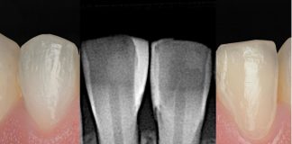
A clinical case by our Community member Dr. Basem Elias
This article and its content are published under the Author’s responsibility as an expression of the Author’s own ideas and practice. Styleitaliano denies any responsibility about the visual and written content of this work.
Interdental papilla loss resulting in open gingival embrasures—commonly referred to as black triangles—represents a common aesthetic concern in restorative and periodontal dentistry. Beyond the cosmetic impact, these spaces may contribute to phonetic alterations, food impaction, and plaque accumulation, potentially compromising periodontal health. The etiology of black triangles is multifactorial and may include periodontal attachment loss, triangular tooth morphology, orthodontic movement, aging, and improper restorative contours.
This clinical case presents the aesthetic closure of black triangles using a direct composite injection technique to improve interdental harmony while preserving dental structure, in accordance with the minimally invasive philosophy.

Fig.1
This case report presents a 50-year-old female patient who was referred by her periodontist after successful management of Stage II, Grade B periodontitis in 2023. Following periodontal stabilization, the patient underwent orthodontic treatment with clear aligners, completed last year. Her chief complaint was the presence of black triangles in between the upper and lower anterior teeth, which compromised the aesthetic outcome despite otherwise successful therapy.

Fig.2
Considering the stability of the periodontal condition and the patient’s primary aesthetic concern, a conservative treatment approach was selected. Since less invasive intervention is generally preferable in such cases, a minimally invasive strategy was discussed and agreed upon with the patient. The treatment plan consisted of direct composite additions to modify proximal tooth morphology and relocate the contact areas, aiming to reduce the appearance of black triangles while preserving sound dental structure.

Fig.3
Left view of the smile.

Fig.4
The retracted view allows for evaluation of the interdental spaces.

Fig.5
Absolute isolation with a rubber dam was achieved using small holes and proper spacing, along with correct inversion to ensure optimal cervical sealing. Biofilm was subsequently removed using sodium bicarbonate powder (Soft 35 μm) delivered with the Aquacare air abrasion system, providing controlled and effective cleaning of the tooth surfaces.

Fig.6
Proper isolation and rubberdam inversion were combined with anatomically shaped Bow anterior matrices (Tor VM), which will help guide the restorative contour. Treatment began with the two maxillary central incisors, while the adjacent lateral incisors were protected using a metal matrix band to prevent unintended bonding and maintain proper proximal contours. The proximal surfaces were then thoroughly cleaned and lightly roughened to enhance adhesion. Air-particle abrasion with 29 μm aluminum oxide (Aquacare) was performed to increase surface energy and promote reliable bonding prior to the adhesive procedure.

Fig.7
A 37% phosphoric acid was selectively applied to the enamel in the proximal area and extended approximately 1 mm beyond the transitional line angles to ensure optimal etching of the bonding substrate. After thorough rinsing and gentle air drying, a two-bottle universal adhesive system (G2 Bond) was applied according to the manufacturer’s instructions prior to composite placement.

Fig.8
One of the main challenges in managing black triangles with direct restorations is creating a correct, hygienic, and smooth proximal emergence profile while maintaining natural tooth morphology and avoiding composite overhangs. To address this challenge, two key elements were implemented.
An injectable universal restorative material (G-ænial Universal Injectable A2) was selected. This thixotropic material provides excellent handling characteristics, allowing controlled placement without oozing, slumping, or uncontrolled flow. In addition, it offers favorable mechanical properties, including high strength, wear resistance, durability, high radiopacity, and very low polymerization shrinkage. The accuracy of the isolation and clamp placement allows precise control of the composite emergence profile and facilitate the creation of a smooth, cleansable proximal surface, leading to predictable aesthetic and functional outcomes.

Fig.9
After light curing the injectable composite in the proximal areas, a packable composite (body shade A2) was applied to the cervical buccal areas. After completing the restorations on the central incisors, the same protocol was repeated for the lateral incisors to achieve harmonious morphology and symmetrical closure of the interdental spaces.

Fig.10
In the following session, the same protocol was repeated for the lower incisors.

Fig.11
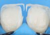
Bow Anterior Matrix shape one.

Fig.12
After composite injection.

Fig.13
Lateral incisor sandblasted and etched.

Fig.14
The restorations were finished and polished using a two-step spiral wheel and cone system. Minor reshaping was performed to refine proximal contours and adjust transitional line angles, allowing better integration with natural tooth morphology and ensuring smooth, hygienic surfaces.

Fig.15
Front view.

Fig.16
Right view.

Fig.17
Left view.

Fig.18
A postoperative radiographic evaluation is essential to verify the integrity of the proximal restorations and ensure the absence of composite overhangs. Radiographic assessment helps confirm proper contouring and adaptation of the restorative material, which is critical for maintaining periodontal health and facilitating adequate interdental hygiene.
Conclusions
Minimally invasive direct composite restorations can provide an effective solution for managing interdental black triangles following periodontal and orthodontic treatment. By carefully modifying proximal morphology and relocating the contact area, it is possible to reduce open gingival embrasures while maintaining periodontal-friendly contours. When performed with proper isolation, matrix systems, and controlled composite placement, this approach offers a predictable, conservative, and highly aesthetic outcome consistent with the minimally invasive philosophy.
Bibliography
- Ramos RQ, Peumans M, Mercelis B, Ahmed MH, Politano G, Lopes GC, Van Meerbeek B. Influence of airborne particle abrasion on dentin bonding effectiveness of a 2-step universal adhesive. J Dent. 2024 May;144:104918.
- Shaalan OO, Abou-Auf E, El Zoghby AF. Clinical evaluation of flowable resin composite versus conventional resin composite in carious and noncarious lesions: Systematic review and meta-analysis. J Conserv Dent. 2017 Nov-Dec;20(6):380-385.
- Manauta J, Salat A. Layers: An atlas of composite resin stratification. 2012. Quintessence Publishing.
- Manauta J, Salat A, Devoto W, Putignano A. Direct composites: The Styleitaliano clinical secrets. 2022. Quintessence Publishing.
- Falacho RI, Melo EA, Marques JA, Ramos JC, Guerra F, Blatz MB. Clinical in-situ evaluation of the effect of rubber dam isolation on bond strength to enamel. J Esthet Restor Dent. 2023 Jan;35(1):48-55.