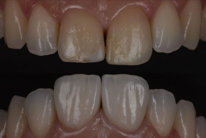
Clinical Case in collaboration with Dr. Daniele Rondoni (DT)
Anterior dark teeth following endodontic therapy represent one of the most demanding esthetic challenges in restorative dentistry. Zirconia veneers offer a predictable and versatile solution, combining strength, precision, and esthetic adaptability. With careful material selection, advanced layering techniques, and meticulous adhesive protocols, clinicians can achieve seamless integration and natural brightness. Veneers thus become more than simple coverings, they serve as transformative restorations that convert compromised teeth into harmonious, radiant smiles.
In this article, we present an anterior esthetic case managed through Zirconia veneering, illustrating how contemporary restorative dentistry can resolve complex discoloration and elevate outcomes into true esthetic harmony.

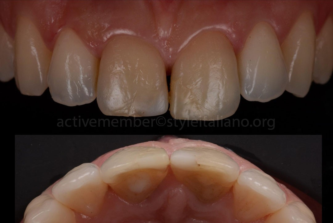
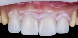
Fig.1
A patient presented to our clinic with an anterior esthetic concern, primarily involving discoloration of the maxillary central incisors. The discoloration was associated with previous endodontic treatment and the presence of aged, defective composite restorations. Following a comprehensive clinical and radiographic assessment, a treatment plan was established involving Zirconia veneers for the maxillary central incisors, after all treatment options were thoroughly reviewed with the patient.
An occlusal evaluation revealed that the central incisors were positioned in lingual version relative to the lateral incisors, allowing for an additive approach with minimal tooth preparation, thereby preserving sound tooth structure.
The esthetic complexity of the case arose from the need to faithfully reproduce the subtle interplay of form, shade, and light transmission within the anterior region to ensure that the final restorations blended imperceptibly with the surrounding natural dentition.
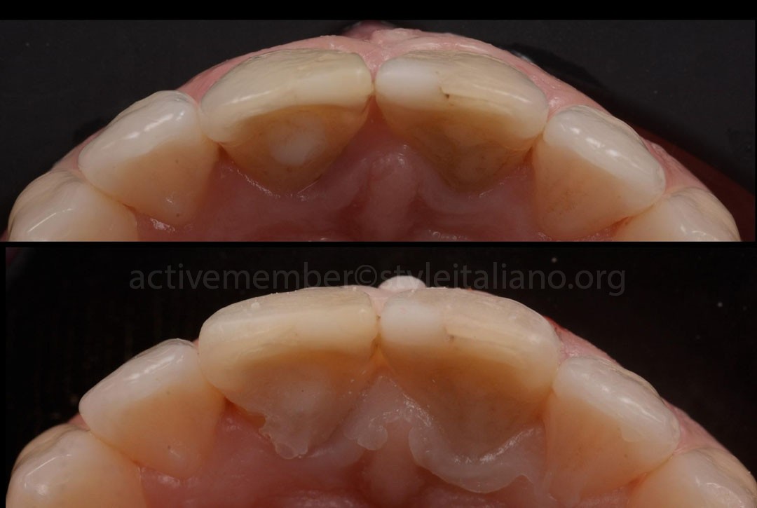
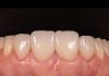
Fig.2
A mock‑up with Filtek Flow Easy Match (Solventum, USA) was fabricated based on the digital smile‑design project, reflecting the planned increase in volume for the maxillary central incisors. From the occlusal perspective, the anterior curvature became clearly evident, illustrating the proposed morphology and guiding the three‑dimensional form of the future veneer restorations.
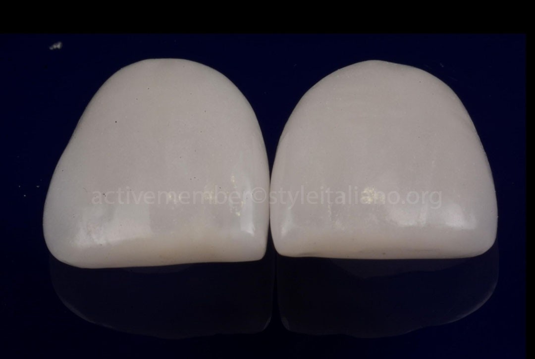
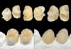
Fig.3
The veneers were produced using a fully digital CAD/CAM workflow, after which the restorations underwent the Zero Cut‑Back protocol, with micro‑layering to refine surface texture and optical characterization.
The zirconia selected for this case, Ezneer (Aidite, Qinhuangdao, China), was conditioned prior to sintering with Biomic LiSi Connect (Aidite, Qinhuangdao, China). This LiSi‑based coating creates a glass‑ceramic–like interface on the zirconia surface, enabling the material to behave more similarly to silica‑containing ceramics during bonding and thereby enhancing its interaction with resin cements.
From a materials‑science standpoint, this pretreatment increases both micromechanical retention and chemical compatibility, ultimately improving adhesive reliability, long‑term performance, and the esthetic continuity of the final restorations.


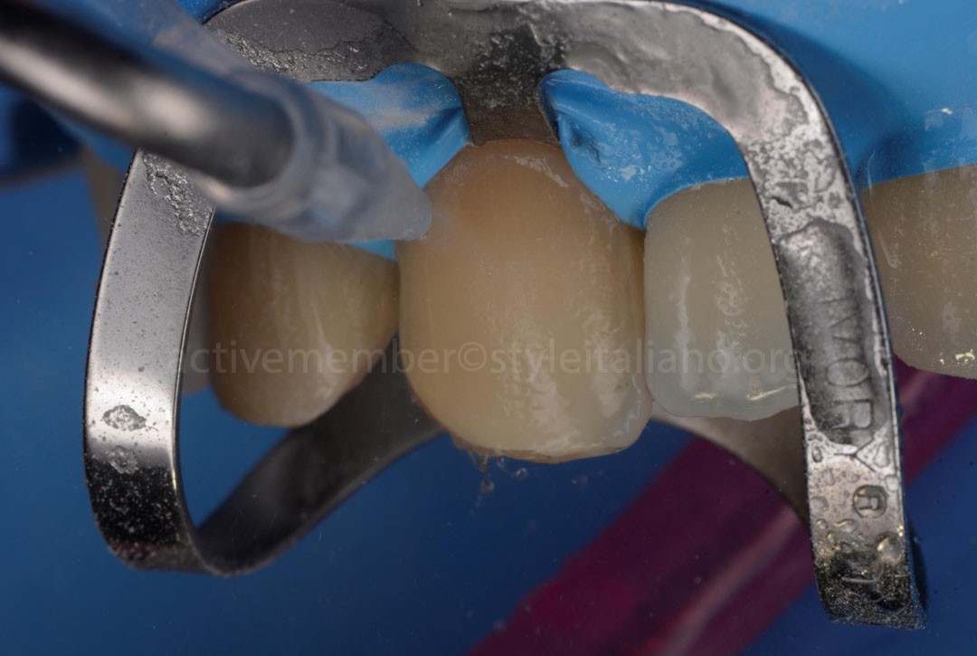
Fig.4
Prior to veneer cementation, the tooth surfaces were air‑abraded using the Acquacare system (Velopex International, London, UK) to eliminate residual temporary cement and to optimize the substrate for adhesive bonding.
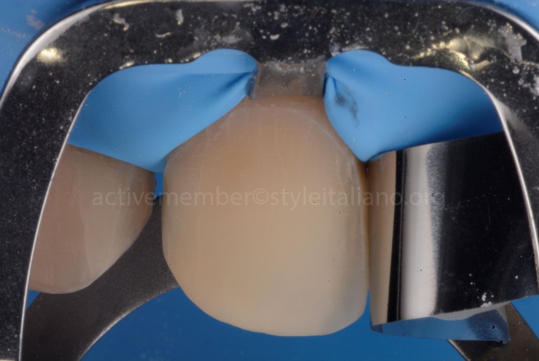
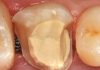
Fig.5
The tooth surface appears clean and properly conditioned following the sandblasting procedure, making it ready for the bonding protocol.
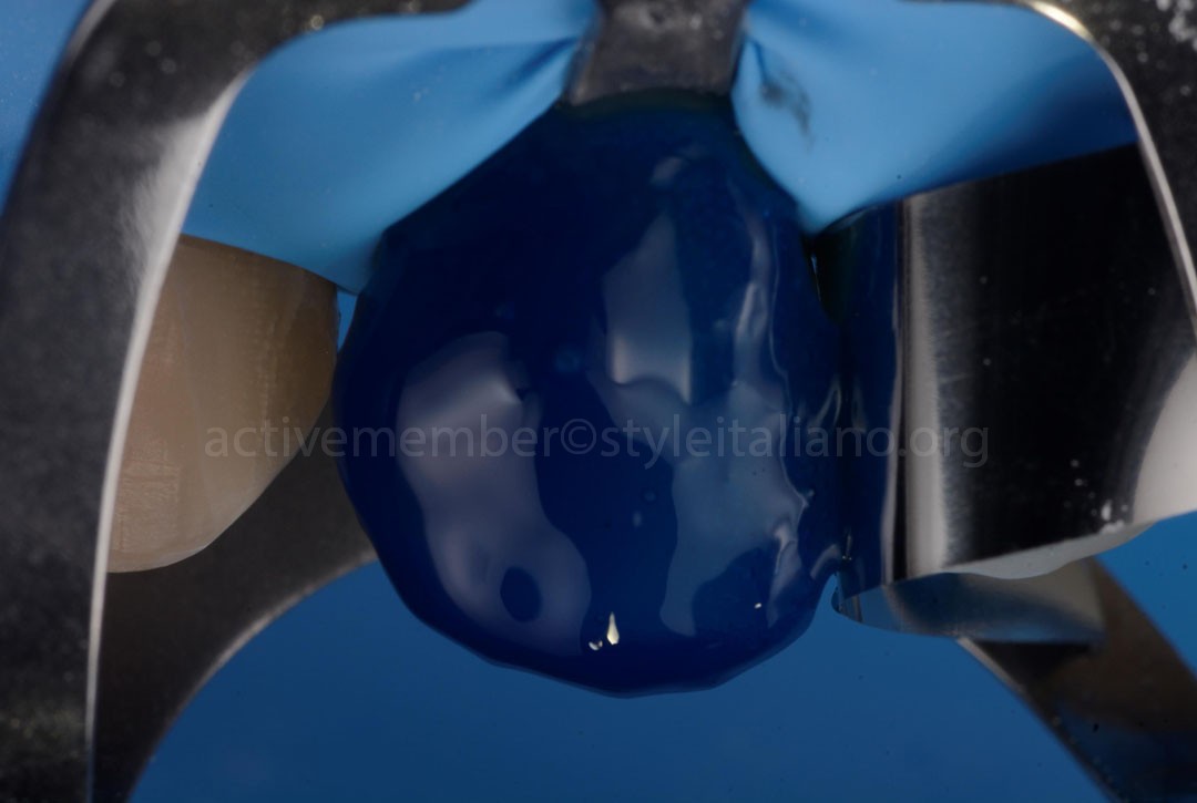
Fig.6
A 37% phosphoric acid etchant was applied to the prepared surface for 15 seconds, with a metallic matrix band placed to protect the adjacent tooth during the procedure.
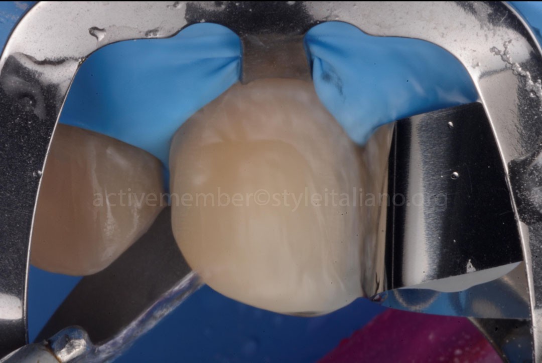
Fig.7
A copious water rinse was performed to thoroughly remove the phosphoric acid, ensuring elimination of both the etchant and any residual reaction products from the tooth surface, thereby promoting optimal adhesive performance.
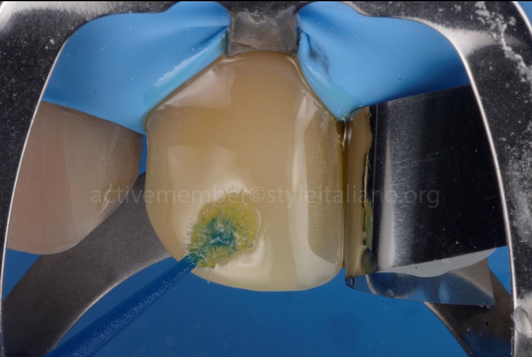
Fig.8
Scotchbond universal plus adhesive (Solventum, USA) was applied with a generous layer on the surface of the centrals, under rubber dam.
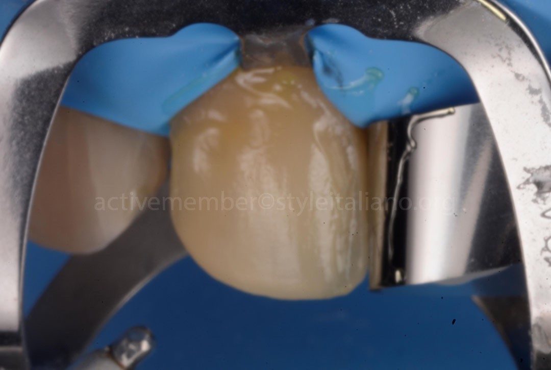
Fig.9
After 10 seconds of gentle air‑drying, the solvent evaporates, and the adhesive transitions from its initial yellowish appearance to a thin, clear, and immobile film, indicating proper readiness for polymerization.
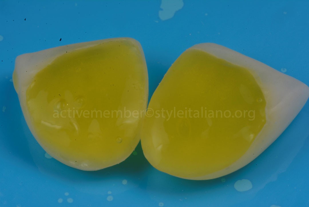
Fig.10
The zirconia surface, previously coated with LiSi Connect and converted into a lithium‑silicate layer, was conditioned with 9.5% hydrofluoric acid for 60 seconds.
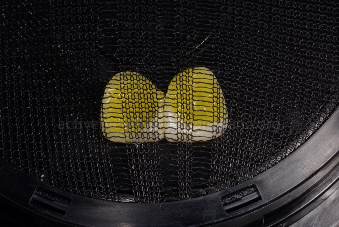
Fig.11
The veneers, still coated with hydrofluoric acid, were placed into the Veneer Me device (Smileline, Switzerland). This system securely stabilizes the restorations and covers them with a protective mesh, allowing the rinsing and subsequent surface treatments to be performed under optimal and highly controlled conditions.
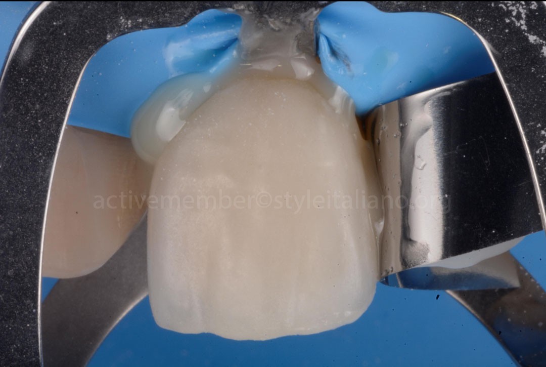
Fig.12
Bonding of the veneers was performed using the RelyX Universal resin cement (Solventum, USA).
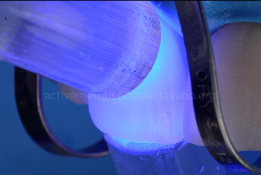
Fig.13
Two light‑curing units were used to polymerize the resin cement, one positioned buccally and the other palatally, to ensure uniform and complete polymerization of the veneers.
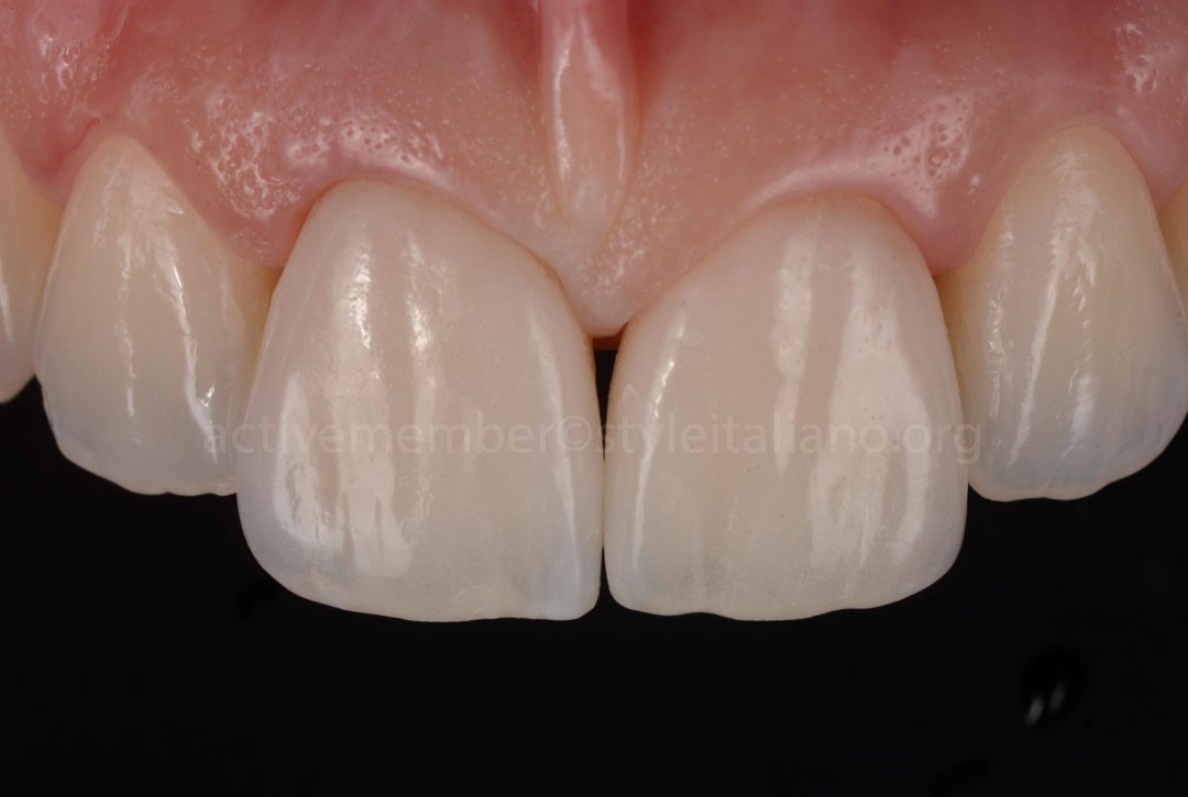
Fig.14
From the buccal perspective, the final outcome demonstrates a seamless integration of the veneers within the anterior esthetic zone.
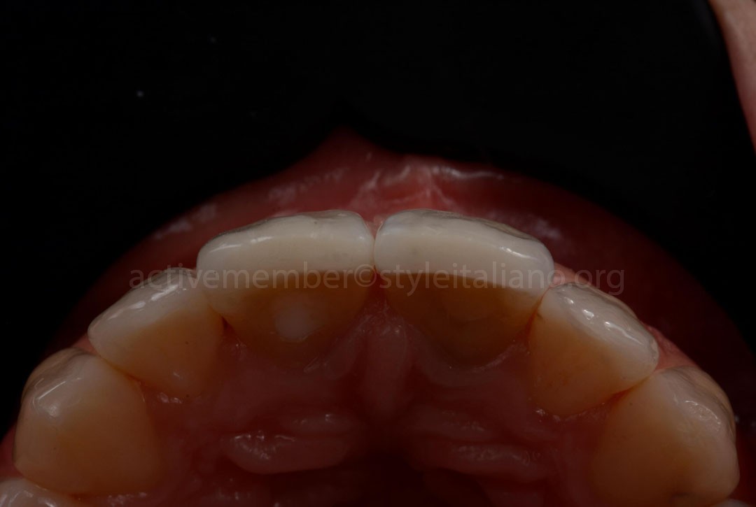
Fig.15
From the occlusal perspective, the final result highlights the newly established buccal contour, with the central incisors now properly aligned in relation to the lateral incisors.
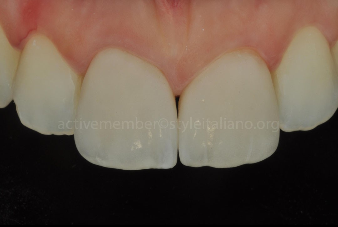
Fig.16
A polarized photograph was captured to evaluate the color integrity of the restorations, confirming both the effective masking of the underlying dark substrate and the harmonious integration with the adjacent teeth.
Note that the gingival margin of tooth 11 was recontoured using LASER therapy to replicate the form and symmetry of the gingiva around tooth 21.
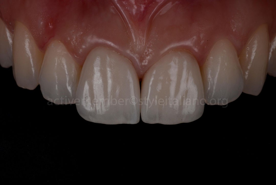
Fig.17
This final low‑exposed photograph highlights the beauty of the Microlayering Zero Cut‑Back technique, showcasing a beautifully defined incisal halo. The secondary anatomy is also clearly visible, contributing to a highly refined and esthetic final outcome.
Conclusions
This clinical case demonstrates how a digitally driven workflow, combined with advanced material protocols, can deliver highly predictable esthetic and functional outcomes. The integration of CAD/CAM‑milled zirconia veneers, enhanced through the Microlayering Zero Cut‑Back technique and the application of LiSi Connect, allowed the restorations to achieve exceptional optical properties and seamless integration within the anterior esthetic zone.
The meticulous surface conditioning, both on the tooth substrate and the lithium‑silicate, coated zirconia, ensured optimal adhesive performance and long‑term stability. Careful execution of each clinical step, from isolation and etching to controlled cementation and polymerization, contributed to a result that is not only esthetically harmonious but also biomechanically sound.
Overall, this case highlights the value of combining modern digital dentistry, innovative material science, and precise adhesive protocols to achieve restorations that meet the highest standards of esthetics, durability, and patient satisfaction.
Bibliography
- El Feghaly, A., Nasr, E., Lukomska-Szymańska, M., Zmysłowska-Polakowska, E., Najjar, G., Harouni, R., Bourgi, R., & Hardan, L. (2026). Effect of different surface treatments on the bond strength of zirconia to resin cement. An in vitro study. Frontiers in Dental Medicine, 7.
- Walter Devoto. When Crowns and Veneers Converge to Restore Esthetics. www.Styleitaliano.org
- Walter Devoto. Beyond Straight Teeth: Zirconia Veneers as the Final Touch in Orthodontic Esthetics. www.styleitaliano.org