Restoring an endodontically retreated posterior tooth with extensive coronal tissue loss represents a significant clinical challenge. In conventional restorative approaches, the presence of an adequate ferrule is considered beneficial for improving fracture resistance and long-term prognosis; however, obtaining a circumferential ferrule may require additional removal of sound tissues or surgical crown lengthening. In maxillary molars, crown lengthening may be particularly unfavorable when the residual tooth structure is close to the furcation area, potentially compromising periodontal support and tooth prognosis.
In selected cases, an endocrown may represent a conservative alternative to a post-and-core supported full-coverage crown. This adhesive monoblock restoration uses the pulp chamber for macromechanical retention and the remaining coronal margins for adhesive stabilization, avoiding further radicular preparation and preserving residual pericervical dentin. Current evidence supports the use of endocrowns particularly in extensively damaged endodontically treated molars, with clinical outcomes comparable to conventional crowns in appropriately selected cases, although long-term high-quality clinical evidence remains limited.

Fig.1
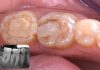
The patient presented with a coronal fracture of an old composite restoration on the maxillary second premolar, while the adjacent maxillary first premolar exhibited an incongruous composite restoration. Both teeth were asymptomatic at the time of examination. Radiographic evaluation revealed inadequate previous root canal treatment of the maxillary second premolar, restored with a metallic Dentatus post. Endodontic retreatment of the second premolar and replacement of both defective coronal restorations were therefore planned. Retreatment was performed using an instrument with a .04 taper to preserve pericervical dentin and adopt a conservative approach in the coronal and middle thirds of the root canal, while allowing appropriate preparation of the previously untreated apical third.

Fig.2
Following endodontic retreatment, the maxillary second premolar presented extensive loss of residual tooth structure. As obtaining an adequate ferrule would have required a more invasive crown-lengthening procedure, an endocrown was selected as the definitive restorative approach. This adhesive restoration exploited the pulp chamber for retention while preserving residual pericervical dentin and periodontal tissues. Conversely, the adjacent first premolar retained sufficient coronal structure to permit a more conservative cuspal-coverage restoration and was therefore prepared for an overlay.

Fig.3
The old restoration on the maxillary first premolar and the provisional restoration on the maxillary second premolar were removed. Both teeth were carefully cleaned, eliminating residual carious tissue and unsuitable restorative substrates. In the second premolar, the coronal portion of the gutta-percha was removed slightly apical to the cemento-enamel junction to create adequate space within the pulp chamber for the endocrown restoration and to prevent potential aesthetic issues related to the dark appearance of the endodontic filling material.

Fig.4
After completion of cavity preparation, immediate dentin sealing (IDS) was performed on both teeth to protect the freshly exposed dentin and optimize the adhesive interface before the indirect restorative procedures. The internal build-up and cavity optimization were subsequently carried out using a flowable bulk-fill composite, SimpliShade™ Bulk Fill Flow (Kerr, California, USA), allowing the restorative substrates to be regularized and prepared for the subsequent endocrown on the maxillary second premolar and overlay on the adjacent first premolar.


Fig.5
The maxillary second premolar was prepared for an endocrown, preserving the remaining enamel to optimize adhesive bonding and maintain a conservative restorative design. The maxillary first premolar was prepared for an overlay with cuspal coverage.

Fig.6
After removal of the rubber dam, two retraction cords were placed to displace the gingival tissues and clearly expose the preparation margins, allowing accurate recording of the finish lines during the subsequent conventional impression-taking procedure.

Fig.7
The model highlights the preparation design of the endocrown on the maxillary second premolar and the overlay on the adjacent first premolar, with clear visualization of the preparation margins and the conservative configuration of both indirect restorations.

Fig.8
The two definitive indirect restorations, consisting of an endocrown for the maxillary second premolar and an overlay for the adjacent first premolar, were fabricated in lithium disilicate and were ready for clinical try-in and subsequent adhesive cementation.

Fig.9
Provisional restorations were fabricated in Telio to protect the prepared teeth during the laboratory phase and to maintain their position within the dental arch, preventing potential tooth movement before definitive adhesive cementation.

Fig.10
The provisional restorations were removed, and rubber dam isolation was performed to achieve optimal moisture control and provide a clean, stable operative field for the subsequent adhesive cementation of the endocrown and overlay restorations.

Fig.11
The prepared tooth surfaces were sandblasted to remove remnants of the provisional restorative material and any contaminants, ensuring clean substrates before the adhesive cementation procedure.

Fig.12
Considering the lithium disilicate composition of both indirect restorations, the intaglio surfaces were conditioned for adhesive cementation. Starting with the endocrown restoration, a 10% hydrofluoric acid gel was applied for 20 seconds, followed by thorough rinsing. Subsequently, 37% phosphoric acid was applied as a cleaning step, instead of ultrasonic cleaning, to remove residual contaminants and reaction products from the internal surfaces before the subsequent adhesive procedures.

Fig.13
A silane primer was actively applied to the entire internal surface of the restorations using a rubbing motion, to promote chemical coupling between the lithium disilicate ceramic and the subsequent resin cementation materials.

Fig.14
Selective enamel etching was performed with phosphoric acid before the application of the universal adhesive.

Fig.15
Following etching application, OptiBond™ Universal 360 adhesive (Kerr, California, USA) was actively applied to the entire internal surface of the lithium disilicate restoration, according to the manufacturer’s instructions.


Fig.16
The universal adhesive was also actively applied to the tooth surfaces using a rubbing and scrubbing motion, ensuring thorough coverage of both enamel and dentin before adhesive cementation.

Fig.17
Maxcem Elite™ Chroma resin cement (Kerr, California, USA) was used for adhesive cementation. During light polymerization, the cement changes color from pink to white, enabling the clinician to visually monitor the progression of polymerization and facilitate the removal of excess cement at the appropriate stage.


Fig.18
The chromatic transition of the resin cement facilitated the identification and removal of excess material. Residual cement was carefully removed using a fine-tipped instrument, such as the LM-Arte Fissura instrument (LM-Dental, Parainen, Finland), and a brush before final polymerization.

Fig.19
To complete polymerization, glycerin gel was applied over the restoration margins to prevent oxygen-inhibited polymerization and ensure complete curing of the resin luting agent at the restoration interface.

Fig.20
The same adhesive cementation protocol was subsequently performed for the lithium disilicate overlay on the first premolar, following the previously described steps.

Fig.21
Final appearance of the restorations after careful removal of the cement excesses and completion of the final polymerization procedure.

Fig.22
Immediate postoperative appearance of the restorations after rubber dam removal, showing their anatomical integration and harmonious aesthetic outcome.

Fig.23
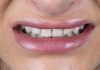
Final clinical appearance of the restorations at the one-month follow-up, showing satisfactory aesthetic integration, preserved marginal adaptation, and healthy surrounding soft tissues.

Fig.24
Radiographic evolution of the clinical case, showing the initial condition, the outcome following endodontic retreatment of the second premolar, and the final appearance after placement of the definitive adhesive restorations.
Conclusions
This clinical case illustrates how an endocrown-based restorative workflow can represent a conservative and effective treatment option for an endodontically retreated posterior tooth with significant coronal tissue loss. By exploiting the pulp chamber for macromechanical retention and combining it with adhesive bonding, the endocrown allowed the tooth to be restored without additional root canal preparation for post placement, thereby preserving the remaining dental structure.
In the present case, the integration of a lithium disilicate endocrown on the second premolar with an adhesive overlay on the adjacent first premolar enabled the recovery of function, anatomy, and aesthetics through a minimally invasive approach. Careful case selection, adequate isolation, precise preparation design, appropriate conditioning of both tooth and ceramic surfaces, and controlled adhesive cementation were essential steps for achieving a satisfactory immediate outcome and stable short-term follow-up.
Although conclusions from a single clinical case must be interpreted cautiously, this workflow may be particularly useful in structurally compromised endodontically treated posterior teeth when sufficient adhesive substrate and favorable occlusal conditions are present. In selected cases, an endocrown may provide a conservative alternative to more invasive restorative approaches, potentially avoiding additional root canal preparation for post-and-core procedures and limiting the need for surgical crown lengthening, thereby preserving the remaining tooth structure and periodontal architecture.
Bibliography
- Barallat L, Arregui M, Fernandez-Villar S, Paniagua B, Pascual-La Rocca A. Fracture Resistance in Non-Vital Teeth: Absence of Interproximal Ferrule and Influence of Preparation Depth in CAD/CAM Endocrown Overlays-An In Vitro Study. Materials (Basel). 2022 Jan 7;15(2):436. doi: 10.3390/ma15020436. PMID: 35057154; PMCID: PMC8778771.
- Dietschi D., Duc O., Krejci I., Sadan A. Biomechanical considerations for the restorations of endodontically treated teeth: A systematic review of the literature-part I. Composition and micro and macrostructure alterations. Quintessence Int. 2007;38:733–743.
- Miluna-Meldere S, Springe B, Papia E. Comparative Analysis of Fracture Resistance in Endodontically Treated Molars Using Lithium Disilicate and Composite Overlays Versus Endocrowns. Int J Dent. 2025 Oct 13;2025:5061799. doi: 10.1155/ijod/5061799. PMID: 41122308; PMCID: PMC12537161.
- Cortellini P, Cortellini S, Bonaccini D, Stalpers G, Mollo A, Buti J. Treatment of Teeth with Insufficient Clinical Crowns. Long-Term Clinical Outcomes of a Minimally Invasive Crown Lengthening Approach: A Retrospective Analysis. Int J Periodontics Restorative Dent. 2025 Jan 24;45(1):46-57. doi: 10.11607/prd.6992. PMID: 37939275.
- de Araújo LNM, Dos Santos MT, Moura DMD, de Assunção E Souza RO, Gurgel BCV. Influence of crown-lengthening surgery on teeth rehabilitated with ceramic single crowns: A 12-month prospective clinical study. J Dent. 2024 Aug;147:105125. doi: 10.1016/j.jdent.2024.105125. Epub 2024 Jun 12. PMID: 38876251.