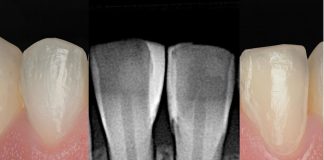
A clinical case by our Community member Dr. Mustafa Yasir
This article and its content are published under the Author’s responsibility as an expression of the Author’s own ideas and practice. Styleitaliano denies any responsibility about the visual and written content of this work.
Natural anterior teeth are defined by more than their shape and shade. Their beauty arises from the subtle interaction of proportion, surface texture, translucency, and light. When tooth structure is severely compromised, the challenge is not to create a perfect restoration, but to recreate a believable tooth within a biologically respectful framework. In this context, direct composite rehabilitation becomes a conservative approach to reconstruction, in which anatomy and optical properties must work together to restore harmony while preserving as much natural tooth structure as possible.

Fig.1
Initial situation of a 17-year-old patient with severely compromised anterior teeth, extensive structural loss, altered proportions, and disrupted smile continuity. The primary challenge was to restore anterior esthetics through a conservative approach while preserving the remaining tooth structure and respecting the long-term biological prognosis.

Fig.2
A disclosing agent transformed the invisible into a diagnostic map by revealing plaque accumulation around the compromised anterior teeth. Before rebuilding esthetics, the biological environment must first be understood, controlled, and respected.

Fig.3
Guided Biofilm Therapy (GBT) with EMS was performed to remove the disclosed biofilm and establish a clean, biologically controlled operating field. Before restoring form and light, the substrate must first be stable, clean, and ready for predictable adhesive procedures. After biofilm control, diode laser gingivectomy was performed to refine the gingival architecture and correct cervical asymmetries. In anterior rehabilitation, the gingiva is not a background, but the biological framework that defines proportion, emergence profile, and overall smile harmony.

Fig.4
Rubber dam isolation, 29 μm aluminum oxide air-abrasion, and selective enamel etching were performed to optimize the adhesive substrate. This protocol enhances surface energy, micromechanical retention, and marginal predictability during direct anterior composite rehabilitation.

Fig.5
A digital mock-up was used to fabricate a silicone index, providing a precise clinical guide for palatal shell fabrication, incisal edge positioning, and final tooth proportions throughout the direct composite build-up.

Fig.6
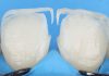
Clear enamel composite was applied through the silicone index derived from the digital mock-up to establish the palatal shell while guiding incisal edge position and final tooth proportions.

Fig.7
The palatal shell was established as the first anatomical reference, defining the incisal edge position, palatal contour, and internal space for the subsequent layering sequence.

Fig.8
Matrices were used to reconstruct the proximal walls and define the contact areas, allowing better control of interdental contour, emergence profile, and final tooth anatomy.

Fig.9
After minimal cut-back and surface reconditioning, the restorations were ready to receive the subsequent composite layers. Controlled space was maintained for optical layering while preserving the previously established anatomy and tooth proportions.

Fig.10
A 1:1 blend of MD and LD was used for the dentin core to control opacity and chroma. BL shade was used to build the proximal walls and define the optical framework of the restoration. A1 body shade shaped the mamelons, blue and ochre tints created internal optical effects, and D1 was added incisally to reproduce the halo effect.

Fig.11
WE shade was applied as the final enamel layer, with controlled adaptation to define the buccal contour, line angles, and surface anatomy before finishing and polishing.

Fig.12
The lateral incisor was restored with the same layering technique, with adjusted mamelon morphology to respect its natural optical behavior.

Fig.13
Final WE enamel layer before finishing and polishing, with facial contour and line angles ready for final refinement.

Fig.14
One-week follow-up showing healthy gingival healing and stable soft-tissue response.

Fig.15
Another view of the final result at the one-week follow-up.

Fig.16
Before and after comparison showing restored proportions, smile line continuity, and natural color harmony. The success of anterior rehabilitation lies in making structure, gingiva, and light read as one balanced composition.
Conclusions
In young patients, direct composite rehabilitation remains one of the most conservative treatment options for anterior rehabilitation. It restores lost tooth structure, corrects tooth contours, and improves the tooth–gingiva relationship without the biological cost associated with fixed prosthetic alternatives.
By re-establishing proper emergence profiles, contact areas, and tooth proportions, direct composite restorations can support gingival health while restoring smile harmony. In this case, treatment provided immediate esthetic and functional improvement while preserving future restorative options for the patient.
At this age, the best treatment is not always the most invasive one, but the one that restores confidence while respecting biological principles.
Bibliography
- Manauta J, Salat A. Layers: An Atlas of Composite Resin Stratification. Quintessence Publishing; 2012.
- Hardan L, Bourgi R, Kharouf N, Mancino D, Zarow M, Jakubowicz N, Haikel Y, Cuevas-Suárez CE. Bond strength of universal adhesives to dentin: A systematic review and meta-analysis. Polymers. 2021 Mar 7;13(5):814.
- Devoto W, Saracinelli M, Manauta J. Composite in everyday practice: how to choose the right material and simplify application techniques in the anterior teeth. Eur J Esthet Dent. 2010 Mar 1;5(1):102-24.
- Dietschi D, Fahl N Jr. Shading concepts and layering techniques to master direct anterior composite restorations: an update. Br Dent J. 2016 Dec 16;221(12):765-71.
- VILLAROEL M, FAHL N, DE SOUSA AM, DE OLIVERA OB Jr. Direct esthetic restorations based on translucency and opacity of composite resins. J Esthet Restor Dent 2011;23(2):73-87.