A clinical case by our Community member Dr. Prjeen Bazeed
This article and its content are published under the Author’s responsibility as an expression of the Author’s own ideas and practice. Styleitaliano denies any responsibility about the visual and written content of this work.
Anterior rehabilitation with direct composite can be challenging when different clinical conditions coexist, such as defective restorations, carious lesions, loss of tooth structure, and esthetic discrepancies. In these situations, treatment should address not only the reconstruction of the compromised tooth structure, but also the optical characteristics required to reproduce a natural appearance.
Achieving this result depends on several factors, including anatomy, material selection, layering, opacity, translucency, and internal characterization. This article presents a comprehensive anterior rehabilitation with direct composite, combining structural reconstruction with optical characterization to restore the anterior region.

Fig.1
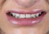
A young female patient presented to the clinic seeking to improve the appearance of her smile. Her main concern was the old composite restorations in the anterior region, associated with color discrepancies and irregular tooth shape.

Fig.2
The retracted intraoral view showed the condition of the existing anterior restorations more clearly, with irregular contours and differences in tooth shape and proportions across the anterior segment.

Fig.3
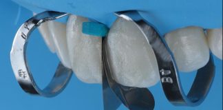
Rubber dam isolation was combined with retraction cord to provide adequate gingival retraction, improve visibility, and maintain proper isolation of the operative field.

Fig.4
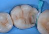
After removal of the old composite restorations and carious tissue, the tooth surfaces were cleaned and conditioned for the adhesive procedure.

Fig.5
The palatal shell was built freehand using a transparent strip and clear enamel composite, establishing the palatal contour and initial framework for the subsequent layering steps.

Fig.6
The missing tooth structure was restored using OPA2 and OA2 composite shades, re-establishing the basic tooth form before proceeding with the layering procedure.

Fig.7
To achieve the brighter appearance requested by the patient, SW was selected as the dentin shade and applied evenly over the facial surface. Its thickness was gradually reduced toward the incisal area to preserve space for the subsequent characterization step.

Fig.8
Internal characterization was performed using clear, blue, lavender, white, ochre, LCO and MCO tints. The tints were selectively applied to define the mamelon pattern and incisal halo, while reproducing the perception of translucency, opalescence and optical depth in the incisal area.

Fig.9
A final layer of WE enamel composite was applied over the facial surface, covering the internal characterization and completing the final contour of the restorations.

Fig.10
Final result after finishing and polishing, showing the completed anterior restorations with refined morphology, surface texture, and integration within the smile.

Fig.11
Before-and-after smile view following anterior rehabilitation with direct composite restorations.

Fig.12
Before-and-after retracted intraoral views.
Conclusions
This case shows how direct composite can be used to manage both structural and esthetic challenges in the anterior region. A controlled layering approach, together with the selective use of tints for incisal characterization, helped reproduce the desired optical effects while maintaining a conservative treatment approach. Careful finishing and polishing completed the final result.
Bibliography
- Devoto W, Saracinelli M, Manauta J. Composite in everyday practice: how to choose the right material and simplify application techniques in the anterior teeth. Eur J Esthet Dent. 2010;5(1):102–124.
- Dietschi D, Fahl N Jr. Shading concepts and layering techniques to master direct anterior composite restorations: an update. Br Dent J. 2016;221(12):765–771.
- Villarroel M, Fahl N Jr, De Sousa AM, De Oliveira OB Jr. Direct esthetic restorations based on translucency and opacity of composite resins. J Esthet Restor Dent. 2011;23(2):73–87.