In clinical implant practice, it is often necessary to apply bone regeneration procedures in cases of horizontal or vertical atrophy of the alveolar process—sometimes in conjunction with maxillary sinus grafting.
Consequently, practitioners must be able to select the surgical procedure best suited to the specific clinical case, as well as the appropriate biomaterials.
Furthermore, allowing sufficient time for biological healing is crucial for achieving complete regeneration of the defect.


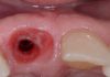
Fig.1
Clinical situation of an edentulous area distal to the first upper premolar with an horizontal hard and soft tissues defect.

Fig.2
Ortopantomography that highlights the insufficient vertical bone quantity with the need to proceed with a crestal sinus floor elevation simultaneously with the insertion of the implants.

Fig.3
Implants inserted after the crestal sinus elevation with the presence of an horizontal bone defect.

Fig.4
Resolution of the horizontal defect by the use of eterologuos bone granules produced with a thermal treatment at high degrees and a resorbable pericardium membrane fixed with pins.

Fig.5
Final x-ray where is possible to evaluate the crestal sinus elevation and the horizontal bone graft.

Fig.6
After 6 months, during the surgical uncovering of the 2 implants, it is possible to appreciate the healing and maturation of the bone graft.

Fig.7
3 weeks after the uncovering procedure the good soft tissue healing, ready for the intra-oral scanning procedure.

Fig.8
PMMA scan base for the intra-oral scanning procedure.

Fig.9
Final zirconia monolithic screwed crowns.

Fig.10
Clinical image of the soft tissue adaptation of the two screw-retained implants crowns.

Fig.11
Intraoral xray where is possible to appreciate the correct fitting and position of the crowns together with the vertical and horizontal GBR.

Fig.12
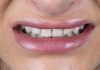
Clinical situation 2 years after the final rehabilitation.
Conclusions
In this clinical case is presented the possibility to combine the resolution of an horizontal and vertical defect of a posterior atrophic maxilla. The sinus crestal elevation allowed us to insert 2 implants in the second posterior quadrant with a simultaneous horizontal and vertical bone regeneration by the use of a resorbable membrane and heterologous bone granules mixed with some autogenous bone particles.
After 6 months the GBR procedure was completed as shown in the clinical image during the implants uncovering.
This procedure allowed us to obtain a healthy emergency profile and a great amount of keratinized tissue around the dental implants. As a consequence, the prostethic rehabilitation with 2 screwed full zirconia crowns, appear more natural because of the regeneration of the hard and soft tissues.
Bibliography
- Sinus membranę elevation by the crestal approach using a novel drilling method. Kim YK, Lee JY, Park JW, Kim SG, Oh JS Implant Dent. 2017,Jun;26(3):351-356.
- Effectiveness of vertical ridge augmentation intervention: a systematic review and meta-analysis. Urban IA, Montero E, Monje A, Sanz-Sánchez I.J Clin Periodontol. 2019 Jun;46 Suppl 21:319-339.
- Limitation and options using resorbable versus non-resorbable membranes for successful guided bone regeneration. Soldatos NK, Stylianou P, Koidou VP, Angelov N, Yukna R, Romanos GE. Quintessence Int. 2017;48(2):131-147.