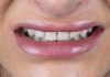
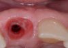
A severely compromised maxillary dentition requiring a staged full-arch rehabilitation. Following extraction, the treatment strategy focused on understanding the residual alveolar anatomy, preserving vascularity, strategically positioning the implants, and allowing adequate healing before second-stage soft-tissue reconstruction. The final rehabilitation demonstrates how diagnosis, biological timing, tissue management, and prosthetically driven planning can transform a challenging clinical situation into a stable functional and esthetic outcome.
Diagnosis. Decisions. Timing. Biology first — prosthetics last.


Fig.1
Case presentation / Initial situation (Diagnosis – Decisions – Timing):
Initial clinical and CBCT assessment showing a severely compromised maxillary dentition.
Aggressive periodontitis bimaxillary condition.
Diagnosis, treatment sequencing and appropriate timing were fundamental to establish a predictable reconstructive strategy.

Fig.2
Post-extraction clinical situation:
Clinical situation following atraumatic extraction of the hopeless maxillary dentition. Careful evaluation of the residual soft-tissue architecture and extraction sockets condition is essential before defining the reconstructive approach.

Fig.3
Soft-tissue management:
Following tooth extraction, meticulous soft-tissue management was performed to preserve the available tissue, optimize vascularization and create favorable conditions for subsequent healing and reconstruction.

Fig.4
Alveolar condition:
Clinical evaluation of the residual alveolar anatomy reveals multiple compromised socket walls and an irregular ridge morphology. The extent and distribution of the defects must guide implant positioning (palatal approach) and the need for staged reconstruction.

Fig.5
Biological consequences after extraction:
Multiple adjacent extraction sockets create a challenging biological environment characterized by disruption of the vascular network, loss of cortical bone and communication between neighboring defects. Preserving the remaining tissue becomes a key component of the treatment strategy.

Fig.6
Strategic implant placement / Palatal approach:
Implants were strategically positioned using the available palatal bone to obtain adequate primary stability while respecting the compromised facial anatomy. Implant positioning was planned according to the definitive prosthetic requirements and the reconstructive limitations of the case.

Fig.7
Healing period (16 weeks):
After an adequate healing period, the surgical site demonstrated complete soft-tissue maturation and favorable tissue volume, providing a stable biological environment for the second surgical stage. Note: the patient used a removable prostheses during this period.

Fig.8
Second-stage surgery (CTG -rotation flap):
Second-stage surgery focused not only on implant exposure but also on soft-tissue optimization. Incision design and flap repositioning were used to preserve and redistribute the available keratinized mucosa around the implants. Surgical extractions of 17, 27 taking CTG from maxillary tuberosity.

Fig.9
Soft-tissue maturation:
Following second-stage healing, a stable peri-implant mucosal architecture was obtained around the restorative components, with favorable tissue thickness and continuity throughout the reconstructed arch.

Fig.10
Keratinized tissue width:
Frontal evaluation demonstrates the resulting band of keratinized mucosa around the implant emergence areas. Soft-tissue quantity and quality were considered integral components of the long-term restorative strategy.

Fig.11
Definitive prosthetic rehabilitation – occlusal view:
Definitive implant-supported rehabilitation after complete tissue maturation. The prosthetic design follows the reconstructed arch while maintaining appropriate access for hygiene and long-term maintenance.

Fig.12
Tissue conformation:
The definitive restoration guides and supports the peri-implant soft tissues, creating a harmonious transition between the prosthetic emergence profile and the reconstructed mucosal architecture.

Fig.13
Final outcome:
Final extraoral result showing restoration of dental function, lip support and smile architecture. The outcome reflects a staged treatment philosophy in which surgical reconstruction, soft-tissue management and prosthetic planning were integrated from the beginning.

Fig.14
A. Initial condition before treatment.
B. Five-month postoperative follow-up at second-stage surgery.
C. Final maxillary implant-supported restoration.
D. Immediate postoperative radiograph following mandibular implant placement.
E. Four-month postoperative follow-up at second-stage surgery.
F. Thirty-month radiographic follow-up under functional loading.
Conclusions
Complex full-arch rehabilitation is not simply about placing implants—it is about managing biology, anatomy, and timing. A staged approach allowed the tissues to heal and mature before definitive reconstruction, while strategic implant positioning and careful soft-tissue management created a stable foundation for the final prosthetic rehabilitation. Respecting biology at every stage remains one of the key factors for achieving predictable functional and esthetic outcomes.
Bibliography
- Gamborena I, Sasaki Y, Blatz MB. Updated clinical and technical protocols for predictable immediate implant placement. J Cosmet Dent. 2020;35:36-51.
- Esquivel J, Gomez-Meda R, Blatz MB. The impact of 3D implant position on emergence profile design. Int J Periodontics Restorative Dent. 2021;41:79-86.
- Esquivel J, Meda R, Villarroel M. Timing implant provisionalization: Decision-making and systematic workflow;J Esthet Restor Dent. 2024;1–10.