In patients with cleft lip and palate, proper management of the residual alveolar cleft is essential to restore the integrity and stability of the maxilla, allow tooth eruption, close the alveolar fistula, and thereby provide support to the lip and nose. Several approaches have been used. Until the 1970s, primary osteoplasty was performed routinely, until Robertson and Rehrmann demonstrated its negative effects on growth. Secondary alveolar bone grafting was subsequently developed and is now well established, following the original procedure described by Boyne and Sands. The most commonly used graft is cancellous bone harvested from the iliac crest, although tibial, mandibular, rib and calvarial bone can also be used. The procedure is usually performed between 9 and 12 years of age, during the mixed dentition phase and before canine eruption, so that the canines can erupt through the grafted site. More recently, other authors have argued that grafting should be performed even earlier, between 4 and 6 years of age and before eruption of the maxillary lateral incisor, allowing the incisor to erupt through the grafted bone. These authors reported better outcomes in terms of residual bone height.
Grafting is classified according to timing. Primary grafting is carried out after lip repair but before palatal repair, whereas secondary grafting is subdivided into early secondary (2-5 years), early mixed dentition (6-8 years), late mixed dentition (9-12 years) and late secondary grafting (after 13 years of age).
Few articles in the literature focus on implant-based treatment of the cleft. Landes demonstrated that the probability of implant success in cleft patients is very similar to that of implants placed after traumatic tooth loss. Oral-health-related quality of life in cleft patients is also similar to that of non-cleft patients.
Matsui et al. emphasized that sufficient bone availability is crucial for successful implant treatment in these patients. Re-augmentation of the cleft jaw (tertiary grafting) is therefore performed with an autogenous iliac crest graft, either three to four months before implant placement or simultaneously with it. Implant therapy is a viable option, but particular attention must be paid to the bone volume required for primary implant stability. Moreover, this complex procedure requires additional soft tissue management. This report describes the minimally invasive implant-supported treatment of a cleft after failed tertiary grafting, with a special focus on hard and soft tissue management, a topic that, to our knowledge, has not previously been described in the literature.


Fig.1
This case presents a challenging clinical scenario — a congenital cleft defect with a complicated history. The patient had previously undergone bone grafting to restore the alveolar ridge, but unfortunately experienced graft exposure, fistula formation, and partial bone necrosis — complications that demanded a comprehensive re-treatment approach.
The Challenge:
- Exposed bone graft with compromised integration
- Persistent fistula connecting oral cavities
- Partial graft necrosis affecting aesthetic and functional outcomes
- Need for complete rehabilitation while recreating hard and soft tissue integrity
Our Solution:
A carefully planned surgical protocol combining advanced grafting techniques, meticulous soft tissue management, strategic implant positioning and prosthetic management to restore both aesthetic harmony and functional excellence.

Fig.2
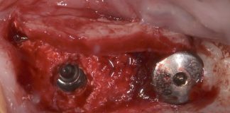
The patient presented with a failed tertiary graft, performed about 1 year earlier with bone harvested from the retromolar region. Radiographic examination showed two osteosynthesis screws retaining residual graft material, with necrosis and an osteolytic area around the screws. The screws and the necrotic bone were removed, and the mucosal fistula was closed during the same surgery. The patient also presented with loss of the mesial papilla of tooth 2.1 and a Miller class III gingival recession.

Fig.3
At 2 months, the mucosa had healed completely, with full coverage of the jawbone. The mucosa was thinner at the site of the previous fistula, but a marked buccolingual and apico-coronal defect and gingival recession persisted. In addition to correcting the defect, the patient asked for an improvement in the esthetics of her smile without orthodontic treatment.
Before planning the prosthetically guided reconstruction, the esthetic and functional parameters relevant to the treatment goal were analyzed.
So it was organised a preview of the potential esthetic outcome using a software of esthetic simulation.

Fig.4
By the use of the simulation software it was decided to involved the adjacent teeth to obtain a correct occlusal and esthetic plane.

Fig.5
An accurate analysis of the digital models is essential for a correct diagnostic wax up. In order to optimize the prosthetic spaces, shapes and volumes it would be necessary to involve the adjacent teeth (#1.1, #2.1 and #2.3).

Fig.6
Matching the digital impression (STL file) with the CBCT Dicom files, made it possible to evaluate the extent of the defect in relation to the soft tissues and the prosthesis to organize a mini invasive hard tissue reconstruction.
The way will be:
1st Buccopalatal reconstruction using heterologous Bone lamina technique associated with autologous particulate bone;
2nd Prosthetical guided implant placement and connective tissue graft
3rd Provisionalization and tissue conditioning
4th Final restoration

Fig.7
Alveolar ridge was reconstructed using a soft heterologous bone lamina, and the gap was filled with autologous bone chips harvested from the mandibular ramus.

Fig.8
Particulate autologous bone placed inside the space between the heterologous bone lamina and the palatal mucosa.

Fig.9
At 6 months, healing was uneventful. Clinically, the regenerated tissues showed a favorable trend, with vertical tissue levels well maintained. The mesial papilla of tooth 2.3 was stable, whereas papillary support at the distal aspect of tooth 2.1 remained insufficient. Hard tissue regeneration was minimal but adequate to accommodate a prosthetically guided implant.

Fig.10
Prosthetically guided implant placement at 6 months after regeneration. The implant was 3 mm in diameter, placed 1 mm subcrestally.

Fig.11
A small-diameter healing abutment was placed, covered with a connective tissue graft (SCTG) harvested from the palate sutured to the base of the anatomical papillae at CEJ level.

Fig.12
Soft tissue healing at 4 months.

Fig.13
A direct mock-up was used to evaluate esthetics and function. It also served as a guide for the veneers on teeth 1.1, 2.1 and 2.3, and for the temporary screw-retained crown on tooth 2.2.

Fig.14
Tooth preparation and customized impression coping for the final impression.

Fig.15
The lithium disilicate veneers and the screw-retained ceramic zirconia crown.

Fig.16
Outcome at 10 years.

Fig.17
At delivery and at 10 years. Respecting the contact point between teeth 2.1 and 2.2 allowed the pseudopapilla to fill the cervical embrasure completely, eliminating any black triangle.

Fig.18
At implant placement, the marginal bone was barely appreciable, and at final prosthetic loading it was almost imperceptible. At 10 years, however, extensive mineralization of the peri-implant bone is evident, confirming the initial subcrestal positioning and adherence to the biological requirements of the transmucosal pathway.

Fig.19
At the end of treatment, the patient achieved a very pleasing smile that fully met her esthetic expectations.
Conclusions
Managing hard and soft tissues in tertiary cleft palate failures was technically challenging. Guided by biological and clinical principles, we adopted a minimally invasive approach to optimize the surgical, implant and prosthetic phases while preserving esthetics and, above all, function.
Bibliography
- Witsenburg B. The reconstruction of anterior residual bone defects in patients with cleft lip, alveolus and palate a review. J Maxillofac Surg 13: 197e208, 1985.
- Robertson NRE, Jolleys A: Effects of early bone grafting in complete clefts of the lip and palate.Plast Reconstr Surg 42: 414e420,1968.
- Rherm AH, Koberg WR, Koch H: Long term postoperative results of primary and secondary bone grafting in complete clefts of the lips and palate. Cleft Palate J 7: 206e212, 1969.
- Boyne PJ, Sands NR: Secondary bone grafting of residual alveolar and palatal clefts. J Oral Surg 3: 87e92, 1972.
- Borstlap WA, Heidbuchel KL, Freihofer HPM, Kuijpers-Jagtman AM: Early secondary bone grafting of alveolar cleft defects: a comparison between chin and rib grafts. J Craniomaxillofac Surg 18: 201, 1990.
- Lilja J, Kalaaji A, Friede H, Elander A: Combined bone grafting and delayed closure of the hard palate in patients with unilateral cleft lip and palate: facilitation of lateral incisor eruption and evaluation of indicators for timing of the procedure. Cleft Palate Craniofac J 37: 98e105, 2000.
- Talmant JC, Lumineau JP, Rousteau G. [Cleft lip, maxilla and palate treatment by Dr. Talmant’s team in Nantes]. Ann Chir Plast Esthet. 2002 Apr;47(2):116-25.
- Eppley BL. Alveolar cleft bone grafting (Part I): Primary bone grafting. J Oral Maxillofac Surg. 1996 Jan;54(1):74-82.
- Rosenstein SW. Early bone grafting of alveolar cleft deformities. J Oral Maxillofac Surg. 2003 Sep;61(9):1078-81.
- Kazemi A, Stearns JW, Fonseca RJ. Secondary grafting in the alveolar cleft patient. Oral Maxillofac Surg Clin North Am. 2002 Nov;14(4):477-90.
- Landes CA, Bündgen L, Laudemann K, Ghanaati S, Sader R. Patient satisfaction after prosthetic rehabilitation of bone-grafted alveolar clefts with nonsubmerged ITI Straumann dental implants loaded at three months. Cleft Palate Craniofac J. 2012 Sep;49(5):601-8.
- Matsui Y, Ohno K, Nishimura A, Shirota T, Kim S, Miyashita H. Long-term study of dental implants placed into alveolar cleft sites. Cleft Palate Craniofac J. 2007 Jul;44(4):444-7.
- Jansma J, Raghoebar GM, Batenburg RH, Stellingsma C, van Oort RP. Bone grafting of cleft lip and palate patients for placement of endosseous implants. Cleft Palate Craniofac J.