Freehand composite veneering is one of the most versatile yet technique-sensitive procedures in aesthetic dentistry. Clinicians must recreate complex anterior tooth anatomy—such as proximal contours, incisal translucency, surface texture, and internal dentinal structures—without the aid of laboratory guides. Minor inaccuracies during layering and shaping can easily compromise the final aesthetic outcome and increase finishing time.
Recent developments in composite materials, sectional matrices, and anatomically designed modeling instruments have improved the predictability and efficiency of anterior restorations by enhancing control over morphology and optical integration.
This case report presents the aesthetic rehabilitation of a maxillary central incisor using a simplified freehand veneering protocol supported by the Replica Anterior instrument.

Fig.1
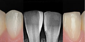
A 30-year-old female patient presented to my dental practice seeking improvement in the aesthetic appearance of her anterior teeth.
At clinical examination, the patient exhibited an unsatisfactory direct restoration on tooth 21, which was compromised in both morphology and chromatic integration with the adjacent dentition. The patient specifically requested replacement of the existing restoration in order to obtain higher aesthetics.

Fig.2
Tooth 21 appeared clinically shorter than the contralateral incisor and presented with visible discoloration. In addition, localized gingival inflammation was observed in the surrounding periodontal tissues, likely associated with the inadequate contour of the existing restoration.

Fig.3
From the palatal perspective, the clinical situation proved even more complex, as tooth 21 was positioned buccally. The patient expressed a desire to correct this malposition through a purely restorative approach, avoiding orthodontic treatment.

Fig.4
The first step consisted of the complete removal of the existing composite restoration. This procedure allowed for a more accurate assessment of the three-dimensional position of tooth 21 and promoted spontaneous healing of the gingival tissues. The previous restoration was clearly overcontoured, extended subgingivally, and exhibited inadequate finishing and polishing, all of which contributed to plaque accumulation and local gingival inflammation.

Fig.5
Subsequently, rubber dam isolation was achieved, maintaining an adequate distance from the gingival margin to enable precise finishing and polishing of the new restoration allowing for healing of the surrounding gingival tissues.

Fig.6
The exposed tooth surface was sandblasted using 29-micron aluminum oxide particles (Aquacare), while the adjacent teeth were carefully protected with a metal matrix strip.

Fig.7
Given that the substrate beneath the removed restoration consisted predominantly of enamel, a total-etch adhesive protocol was selected.

Fig.8
A self-etch bonding agent was actively applied to the prepared enamel surfaces for 20 seconds, gently air-thinned, and subsequently light-cured for 40 seconds.

Fig.9
To remove the B4 clamp at an early stage and gain a wider operative field, the cervical portion was layered first. Due to the need for increased opacity, a dentin shade (UD3, Micerium) was used. An anatomically designed sectional matrix developed by StyleItaliano (Unica Proximal, Polydentia) was placed to build both proximal walls, starting from the distal one. The matrix sits firmly at the level of the papilla and maintains a secure fit within the embrasure, enabling precise and predictable reproduction of proximal anatomy.

Fig.10
A novel modeling instrument was then used to sculpt the composite in the proximal area. The Replica Anterior instrument (LM, Finland), specifically designed for anterior veneering procedures, was used as a single-instrument solution for precise composite manipulation. Owing to its unique design and in combination with the self-polishing properties of the selected sectional matrix, the external surface of the matrix was gently pressed to adapt the composite accurately along both the tooth surface and the matrix profile. For this phase, a dentin shade composite (UD2, Micerium) was applied to recreate natural internal anatomy.

Fig.11
As a result of the appropriate selection of both the sectional matrix and the modeling instrument, the distal wall of tooth 21 closely replicated the morphology of the corresponding surface of tooth 11.

Fig.12
Freehand veneering requires the “box” to be built first. In this clinical case, the Replica Anterior instrument proved extremely precise and effective for both shaping the composite and removing excess material. The incisal edge was also built using a dentin shade composite.

Fig.13
The morphology of the Replica Anterior instrument is characterized by a wavy, anatomically inspired design that facilitates controlled shaping of dentinal mamelons and guides their accurate replication during subsequent enamel layering.
Result as soon as the rubber dam is removed. It is surprising to see how good the blend of composite with the natural tooth is and the natural shapes. Once the optical width of the restored teeth is established, a disc is sufficient to draw the transition lines.

Fig.14
Following completion of the dentin core, a minimal amount of characterization stain may be applied to the incisal margin.

Fig.15
In the final layering phase, an enamel shade composite (UE2, Micerium) was applied in a single, uniform layer.

Fig.16
When transilluminated with a curing light, the composite material exhibited high fluorescence; however, under natural sunlight conditions, the fluorescence closely matched that of the adjacent teeth.

Fig.17
The final result was evaluated, and the patient was scheduled for a short-term follow-up visit to perform any required adjustments.

Fig.18
Following rubber dam removal and gentle subgingival debridement, mild bleeding was observed from the previously inflamed gingival margin.

Fig.19
Final polishing procedures were performed at the follow-up appointment a few days later, and no additional color adjustments were required.

Fig.20
At the 6-month follow-up, the restoration demonstrated excellent stability in terms of color match and surface gloss, although the patient was advised to attend a professional dental hygiene session. The treatment outcome fully met the patient’s expectations regarding shade integration, morphology, and tooth alignment. Of particular clinical relevance was the use of a single dedicated instrument, the Replica Anterior, specifically designed for veneering procedures. Its application—especially in cases involving multiple restorations—allows for a substantial and clinically significant reduction in overall chairside time.

Fig.21
Before and after.
Conclusions
The use of the Replica Anterior instrument significantly simplifies and enhances the predictability of anterior composite restorations. Its wavy, anatomically inspired design enables precise formation of dentinal mamelons and effectively guides their replication during enamel layering, thereby minimizing the need for time-consuming adjustments. versatile modeling instrument, it facilitates accurate composite adaptation along both the tooth surface and the sectional matrix, making sculpting, contouring, and removal of excess material considerably easier—even in complex proximal areas. Combined with sectional matrices, it ensures stable positioning and easy reproduction of the natural anatomy, allowing clinicians to consistently achieve high-level aesthetic outcomes with greater efficiency. Overall, the Replica Anterior streamlines the restorative workflow, making freehand anterior veneering faster, more controlled, and less technique-sensitive.
Bibliography
- Monterubbianesi R, Tosco V, Orillisi G, Grandini S, Orsini G, Putignano A. Surface evaluation of a nano composite after different finishing and polishing systems for anterior and posterior restorations. Microscopy Research & Technique, 2021.
- Bourgi R, Hardan L, Cuevas-Suárez CE, Devoto W, Kassis C, Kharma K, Harouny R, Ashi T, Mancino D, Kharouf N, et al. Effectiveness of Different Application Modalities on the Bond Performance of Four Polymeric Adhesive Systems to Dentin. Polymers2023,15,3924.
- Heintse SD, Rousson V, Hickel R. Clinical effectiveness of anterior restorations – a meta analysis. Dental Materials 2015, 31: 481-495.
- Demarco FF, Collares K, Correa MB, Cenci MS, Moraes RR, Opdam NJ. Should my composite restorations last forever? Why are they failing? Brad Oral Res 2017 Aug 28;31 (suppl 1):e56.
- Manauta J, Salat A, Devoto W, Putignano A. LAYERS 2. Direct composites: the StyleItaliano clinical secrets.
- Demarco FF, Baldissera RA, Madruga FC, Simões RC, Lund RG, Correa MB. Cenci MS. Anterior composite restorations in clinical practice: findings from a survey with general dental practitioners. J Apple Oral Sci. 2013.