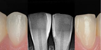
Direct anatomical remodeling in the upper anterior region has become a routine procedure in daily clinical practice. Patients increasingly seek aesthetic solutions for concerns such as alterations in tooth shape or color, diastemas, and dental wear. Conservative, minimally invasive techniques that preserve tooth structure are the ideal approach to address these demands. The goal is to achieve restorative outcomes that are predictable, reproducible, and easy to teach, ensuring consistent clinical success.

Fig.1
The patient presented with multiple defective restorations, noticeable wear, and disproportional anterior teeth requiring comprehensive esthetic rehabilitation.

Fig.2
The image highlights the shape alterations and defective restorations affecting the six upper anterior teeth. The treatment plan focuses on redesigning the individual and collective tooth anatomy to re-establish smile harmony.

Fig.3
The image highlights the shape alterations and defective restorations in the six upper anterior teeth. The treatment plan focuses on redesigning the individual and group anatomy to restore smile harmony.


Fig.4
The image documents the tooth shade and color mapping, which will guide the esthetic planning and restorative process.

Fig.5
The image shows the clinical preparation, including cleaning, isolation, and elimination of defective restorations prior to restorative treatment.

Fig.6
The total-etch technique was performed on the large enamel surfaces to ensure a reliable adhesive pattern. Phosphoric acid 35% was applied for 25 seconds, rinsed, and dried. One Coat 7 Universal (Coltene, Ailstatten, Switzerland, Europe) was rubbed over the entire surface, gently air-thinned for 5 seconds, and polymerized for 10 seconds.

Fig.7
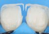
The palatal wall was first built, either freehand or using a silicone key derived from an analog or digital wax-up on a 3D model. Undercuts were then filled, followed by cervical augmentation to establish the emergence profile. Guided by the Polydentia Unica matrix (Polydentia, Mezzovico-Vira, Switzerland), the mesial and distal walls were created. A translucent or achromatic composite (Brilliant Everglow Trans, 27% translucency, Coltene, Ailstatten, Switzerland, Europe) was used for the palatal wall, and a body or intermediate translucency composite (Brilliant Everglow Universal, 21% translucency, Coltene, Ailstatten, Switzerland, Europe) for the cervical, mesial, and distal walls. Each layer was polymerized for 10 seconds.

Fig.8
After the Unica matrix guided the reconstruction of difficult areas, it was removed to complete the vestibular layers. Composite body was added to create mamelons using LM Solo Anterior, LM Fissura (LM-Dental, Parainen, Finland), and flat brushes for precise adaptation. Care was taken to avoid the incorporation of air bubbles.

Fig.9
To naturally reproduce the incisal area, a white pigment (Effect Miris White, Coltene, Ailstatten, Switzerland, Europe) was applied and polymerized to enhance translucency and optical effects.

Fig.10
A final layer of translucent enamel was applied to cover the middle third to the incisal edge, completing the anatomic and optical integration of the restoration.

Fig.11
A final layer of translucent enamel composite was applied to cover the middle third to the incisal edge, completing the restoration’s anatomical and optical integration.

Fig.12
The central incisors were pre-finished, then protected with matrices while the same procedure was applied to the lateral incisors.

Fig.13
The palatal wall, emergence profile, and mesial and distal walls of the lateral incisor are shown.

Fig.14
The central incisors were used as a reference to maintain proper proportions with the laterals and canines. The incisal edge positions, zeniths, embrasure angulations, and contact points were carefully respected to achieve a natural result. After finishing the lateral incisors, the same procedure was applied to the canines.

Fig.15
Medium-grain discs were used to define the basic and primary anatomy, followed by a long conical stone to refine V-shaped depressions and areas within the reflection lines. Polishing was performed with aluminium oxide and diamond rubber discs (Diatech Coltene ShapeGuard), and a high gloss was achieved using the Diashine Lucida System.

Fig.16
Black marks indicate the elevations of the refraction lines, while red marks highlight the V-shaped depressions.

Fig.17
The restorations are completed, showing the elements required for finishing, pre-polishing, polishing, and achieving a high gloss.

Fig.18
Front view in occlusion, showing the completed restorations.

Fig.19
Lateral view showing the finished restorations and the different planes in the cervical, middle, and incisal areas.

Fig.20
Front view in occlusion, showing the completed restorations.

Fig.21
Final situation.
Conclusions
In recent years, advances in materials, matrix systems, instruments, devices, and clinical protocols have enabled dentists to manage many challenging cases directly, conservatively, and efficiently, even when high esthetic demands are required. Simplified protocols for fabricating direct composite resin veneers exemplify this progress. Mastering and consistently applying a precise method allows clinicians to achieve predictable, repeatable, and highly esthetic restorations.
Bibliography
- DEVOTO W. , SARACINELLI M., MANAUTA J. Composite in everyday practice: how to choose the rightmaterial and simplify application techniques in the anterior teeth. The European Journal of Esthetic dentistry. Volumen 3. Número 3. Año 2010.
- DIETSCHI D. Stratification concepts in anterior composite restaurations. J Adhesive Dent 2001. 3: 71 – 80.
- MANAUTA J, SALAT A, PUTIGNANO A, DEVOTO W, PAOLONE G, HARDAN LS. Stratification in anterior teeth using one dentine shade and predefined thickness of enamel. A new concept in composite layering. Parte 2 . Odontostomatol Trop 2014: 37 ( 47): 5-13.
- MANAUTA J, SALAT A, LAYERS An Atlas of composite resin stratification . Quintessence Books, 2012.
- BAZOS P, MAGNE P, Bio-Emulation biomimetically emulating nature utilizing a histoanatomic approach; visual synthetics. The International Jornal esthetic Dentistry. Volume 9. Number 3. Autumn 2014.
- HIGASHI C, GOMEZ JC, KINA S, SCOPIN DE ANDRADE O, HIRATA R, planejamento estetico em dentes anteriores ( Cap 7 ) In: Odontología Estética Planejamento e tecnica ed. Sao Pablo: Artes medicas Ltda. Divisao Odontologica . 2006.
- BLITZ N. Criteria for success in creating beautiful smiles. Oral Health 1997; 87: 38–42.
- VILLAROEL M, FAHL N, DE SOUSA AM, DE OLIVERA OB Jr. Direct esthetic restorations based on translucency and opacity of composite resins. J Esthet Restor Dent 2011;23(2):73-87.