Discoloration of non-vital teeth is a frequent finding, especially after dental trauma. Endodontically treated teeth often undergo intrinsic chromatic changes due to pulp tissue degradation, blood breakdown products, or remnants of endodontic materials. These factors compromise translucency and esthetics, affecting patient confidence.
Internal bleaching is the primary treatment option, aiming to neutralize discoloring compounds within the pulp chamber. Yet, outcomes are sometimes incomplete or short-lived.
In such cases, alternative or combined approaches may be required, including external bleaching or restorative solutions like veneers and crowns. Modern adhesive and minimally invasive techniques allow clinicians to tailor treatment, balancing esthetics, function, and tooth preservation.

Fig.1
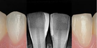
A 36‑year‑old male presents with persistent discoloration of tooth 21.
The tooth had sustained trauma and was treated with root canal therapy 10 years ago.
Two internal bleaching sessions were performed previously at another clinic, resulting in partial shade improvement but leaving noticeable discoloration.
The incomplete response suggests residual intrinsic factors, and further esthetic management is required.

Fig.2
Localized discoloration is evident on tooth 21, presenting a noticeably darker shade compared to the adjacent incisors.
The contrast in color highlights the esthetic discrepancy within the anterior region, drawing attention to the affected tooth and impacting the overall harmony of the smile.

Fig.3
Digital shade evaluation was performed using OptiShade, revealing baseline values of L 71.3, a 4.0, and b 19.6.
The corresponding reference shade was almost 3.5M2, confirming a darker chromatic profile compared to the adjacent teeth.
The patient reported no endodontic symptoms and the Xray revealed a good endodontic treatment.

Fig.4
The adjacent teeth exhibited a 1M1 shade, highlighting a pronounced contrast with tooth 21 and emphasizing the significant color discrepancy.
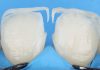
A localized external home bleaching protocol was applied exclusively to tooth 21 in order to prevent over‑whitening of the adjacent teeth.
The treatment utilized a 16% carbamide peroxide (White Dental Beauty, UK) gel delivered through a customized tray with a localized reservoir.
Follow‑up evaluations were scheduled at 2, 4, and 8 weeks to monitor progress and ensure controlled shade improvement.


Fig.5
The treatment was carried out over a total duration of 8 weeks. Shade evolution, recorded with OptiShade (SmileLine, Switzerland), demonstrated a significant improvement: luminosity increased (ΔL +3.8), while both yellow (Δb –8.0) and red (Δa –2.6) chromatic components decreased. These changes resulted in a noticeably closer shade match with the adjacent teeth (1M1), enhancing overall esthetic integration.

Fig.6
A very minimal preparation was performed on tooth 21 to allow masking of the residual discoloration with composite resin.

Fig.7
A final restorative phase was carried out to optimize esthetic integration.
Incremental composite additions were placed on the buccal and interproximal areas of teeth 11 and 21, aiming to mask residual chromatic depth. This approach also allowed for closure of the interdental black triangle, reconstruction of proximal contact, and redistribution of volume to achieve a more harmonious anterior appearance.

Fig.8
A nano‑hybrid light‑cured composite was applied using a minimally invasive approach, without the need for significant tooth preparation.

Fig.9
Shade harmonization with the adjacent teeth was successfully achieved, with a marked reduction in visible discoloration.
The cervical contour and light diffusion were noticeably improved, and no postoperative complications were observed.

Fig.10
This case highlights that even when internal bleaching produces incomplete results, an extended protocol of selective external bleaching can achieve significant shade improvement while avoiding more invasive options such as veneers or crowns. The subsequent composite refinement served as an adjunctive solution, enhancing cervical esthetics, masking residual chromatic shadow, restoring interproximal morphology, and improving overall optical integration. The combined approach of progressive bleaching and targeted conservative restoration ultimately provided a stable, minimally invasive, and cost‑effective outcome, reinforcing the value of conservative strategies in esthetic dentistry.
Conclusions
The combined treatment, consisting of an 8‑week protocol with 16% carbamide peroxide followed by cervical and interproximal composite additions, achieved successful chromatic integration of the previously discolored non‑vital tooth without the need for prosthetic intervention. This approach proved to be minimally invasive, controllable, and repeatable, representing a valid therapeutic option for cases of persistent post‑endodontic discoloration.
Bibliography
- Sara Laface. When a tray solves it all: How to treat dyschromia with non-invasive dentistry. Styleitaliano.org
- Sara Laface. Align, Bleach and Restore (ABR concept) Part II: A gentle path to a naturally beautiful smile. Styleitaliano.org
- Rim Bourgi. Evolution of dental bleaching, from research to clinical evidence. Styleitaliano.org