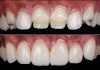
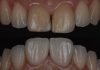
A clinical case by our Community members Dr. Rim Bourgi and Dr. Mohammad Qaddomi
This article and its content are published under the Author’s responsibility as an expression of the Author’s own ideas and practice. Styleitaliano denies any responsibility about the visual and written content of this work.
Digital dentistry has revolutionized the approach to complex cases, incorporating advanced technologies that enhance accuracy, efficiency, and patient satisfaction. By integrating artificial intelligence (AI), digital smile simulation, and sophisticated guide designs, dental professionals can achieve more predictable outcomes, improve patient communication, and streamline workflows in both restorative and surgical procedures.
In this case, a digital approach was used to simplify a complex gingivectomy procedure. The patient presented with challenging gingival architecture, requiring precise planning and execution. The treatment process began with an AI-driven smile simulation, followed by the creation of two digital gingivectomy guides and a milled provisional restoration. These digital tools enabled enhanced visualization, ensured accurate guide placement, and facilitated a more predictable outcome. The digital workflow resulted in a minimally invasive procedure, improved efficiency, and a high-quality mock-up, enhancing both aesthetic and functional results.

Fig.1
In the past, complex and multidisciplinary cases were not commonly managed due to the challenges associated with traditional techniques. However, digital solutions have provided practical, easy, and reproducible alternatives.
A patient presented with a smile that required aesthetic correction. Clinical examination revealed a gummy smile along with suboptimal dental and gingival architecture.
To address this, the problem was approached using advanced knowledge in digital dentistry.
A scan was performed using the Helios 500 from Eighteeth Company (Changzhou, Jiangsu Province, China) to visualize both the upper and lower arches.

Fig.2
The Polygon File Format (PLY) file of the upper jaw was imported into Exocad software (Exocad GmbH, Darmstadt, Germany), revealing increased gingival volume and a non-ideal architecture of both the teeth and gingiva.

Fig.3
Initial buccal view of the maxillary anterior teeth.

Fig.4
Clinical evaluation using the Chu proportion gauge, indicating reduced height of the maxillary central incisor relative to ideal proportions.

Fig.5
Three-dimensional (3D) facial scan of the patient acquired using the MetiSmile scanner (Shining 3D Tech. Co., Ltd., Hangzhou, China), serving as supplementary data for comprehensive digital treatment planning.
Superimposition of the initial intraoral scan and Cone-Beam Computed Tomography (CBCT) data using SmileCloud software (SmileCloud SRL, Timișoara, Romania) to generate a comprehensive 3D smile design integrating dental, skeletal, and soft tissue structures.

Fig.6
3D smile design showcasing the ideal correction of tooth alignment and gingival harmony, with adjustments made for an aesthetically balanced smile. The design includes the refinement of the gingival margin, correction of tooth position, and alignment of the smile zenith curve to ensure optimal facial and dental aesthetics.

Fig.7
An AI–based smile simulation was performed using SmileFy software (SmileFy Inc., Hallandale Beach, FL, USA) by capturing a frontal facial photograph of the patient, which was then integrated with the digital scan to visualize the proposed smile design.

Fig.8
The two-dimensional (2D) smile design, generated by the AI-powered software, clearly illustrates the ‘before’ and ‘after’ outcomes, offering a user-friendly and efficient tool for treatment planning.
Following review of the 2D smile design, the patient agreed to proceed with treatment, and a 3D smile design was subsequently created. So, the 2D smile design provides a rapid and clear visual representation of the potential outcome, allowing both the clinician and patient to quickly assess the aesthetic changes.

Fig.9
Before and after 2D smile design simulation using Smilecloud software (SmileCloud SRL, Timișoara, Romania), showcasing the AI-powered customization based on the patient’s facial features.

Fig.10
Models of the initial and smile design were printed using model resin, ensuring high precision and durability for further evaluation and treatment planning.

Fig.11
After completing the 3D smile design, a guide for the gingivectomy procedure was created using Exocad software (Exocad GmbH, Darmstadt, Germany).

Fig.12
Another guide was designed using CoDiagnostiX software (Straumann, Basal, Switzerland).
The use of two guides offers several advantages:
- Increased Accuracy and Precision: The two guides allow for better alignment and precision during procedures such as gingivectomy in this case. By using different software tools, each guide is optimized for specific aspects of the treatment, ensuring greater overall accuracy.
- Redundancy for Increased Confidence: Using two guides created with different software systems provides an added layer of security. This redundancy ensures that any potential errors or misalignments in one system are compensated for by the other, leading to a more predictable outcome.

Fig.13
Here are the two guides printed on the patient’s initial model. Next, the guides were stored in a dark environment to prevent any potential errors during the gingivectomy procedure and to preserve the dimensional stability of both guides, as previously demonstrated in the literature. Additionally, to maintain precision, guides should be used within 7 days and stored in a dry environment to prevent any alteration in their dimensional stability.

Fig.14
Frontal view of the guide superimposed on the 3D model, demonstrating that the gingival margin is safely positioned away from the underlying bone, confirming the absence of need for a crown lengthening procedure.

Fig.15
Lateral view of the guide superimposition showing a clear distance between the planned gingival incision line and the alveolar crest, ensuring a conservative and biologically safe gingivectomy.

Fig.16
Intraoral positioning of the Exocad-designed guide to assess its fit and adaptability.
Reference lines drawn through the guide to outline the gingival tissue to be removed, confirming alignment with the 3D smile design.

Fig.17
This image shows the gingivectomy procedure performed using a laser and guided by a fully digital workflow. The surgery was carried out by Prof. Karim Corbani.

Fig.18
Lateral view of the surgical guide positioned intraorally during the gingivectomy procedure performed with the Erbium-doped Yttrium Aluminum Garnet (Er:YAG) laser. The image was captured using the Smile Lite MDP2 (Smile Line, Switzerland) at full power in combination with a Samsung S22 Ultra smartphone. For soft, evenly diffused lighting, all six LEDs on each lateral panel and the eight LEDs on the central panel were activated and covered with the white diffuser.

Fig.19
Milled provisional restorations with palatal support, designed according to the finalized 3D digital smile design, were fabricated to ensure optimal fit, enhanced fracture resistance, and reliable aesthetic and functional evaluation.

Fig.20
Occlusal view of the milled computer-aided design/computer-aided manufacturing (CAD/CAM) provisional restorations in the patient’s mouth, showcasing the palatal support and harmonious curvature of the anterior teeth for optimal aesthetic and functional integration. The restorations were fabricated using the Zirkonzahn M1 milling unit (Zirkonzahn, Gais, Germany) and Mono Polymethylmethacrylate (PMMA) discs (98.5 mm × 20 mm, A1 shade) from Aidite (Qinhuangdao Technology Co., Ltd., China). This specialized temporary CAD/CAM material, composed of 99% PMMA, is recognized for its exceptional mechanical strength, flexibility, structural stability, and aesthetic properties, including high gloss and natural translucency. These features make Mono PMMA an ideal choice for long-term provisional restorations that closely mimic natural dentition.

Fig.21
Before and after provisional placement: noticeable aesthetic transformation following full digital workflow and insertion of milled CAD/CAM provisional restorations.
The patient can see the final result, and the mock-up will serve as a guide for preparation.
Conclusions
The integration of digital solutions, including AI for smile simulation and the use of digital guides, significantly simplified the treatment process for a complex gingivectomy case. The use of digital technologies not only enhanced the precision of the gingivectomy procedure but also ensured an optimized fit for the milled mock-up which can serve as a provisional restoration.
This approach highlights the transformative potential of digital dentistry in addressing challenging clinical scenarios, offering both improved patient outcomes and greater efficiency in treatment planning and execution.
Bibliography
- Blatz MB, Chiche G, Bahat O, Roblee R, Coachman C, Heymann HO. Evolution of esthetic dentistry. J Dent Res. 2019;98(12):1294-304.
- Reis IN, Damin GB, Pereira CR, de Alencar Ichigi M, Sant’Anna LO, Spin-Neto R, et al. Digital planning to enhance diagnosis and precision in correcting excessive gingival display in the presence of asymmetrical maxillary position: a case report. Eur J Dent. 2024;18(3):950-6.
- Alrebdi A, Atria P, Teehankee MT, Olcay V, Lopez J, Sampaio CS. Fully digital workflow involving 3D-printed gingivectomy guide and 3D-printed wax-up to restore and reshape a congenitally missing central incisor after orthodontic treatment. Quintessence Int. 2025;56(1).
- Coachman C, Calamita MA, Sesma N. Dynamic documentation of the smile and the 2D/3D digital smile design process. Int J Periodontics Restorative Dent. 2017;37(2):183-93.
- Galbraith A, Doan M, Galbraith T, Abubakr NH. Evaluation of color stability and marginal integrity in provisional restorations: A study of milling, 3D printing and conventional fabrication methods. Dent J. 2025;13(2):189.
- Lee JD, Nguyen O, Lin YC, Luu D, Kim S, Amini A, et al. Facial scanners in dentistry: An overview. Prosthesis. 2022;4(3):664-78.
- Piedra‐Cascón W, Fountain J, Att W, Revilla‐León M. 2D and 3D patient’s representation of simulated restorative esthetic outcomes using different computer‐aided design software programs. J Esthet Restor Dent. 2021;33(1):143-51.
- Ntovas P, Marchand L, Basir B, Kudara Y, Revilla‐Leon M, Att W. Effect of storage conditions and time on the dimensional stability of 3D printed surgical guides: An in-vitro study. Clin Oral Implants Res. 2025;36(1):92-9.
- Ozden YE, Ozden I, Kayahan ZO, Kazazoglu E. The effect of storage conditions and duration on dimensional stability of 3D-printed endoguides. BMC Oral Health. 2025;25(1):641.
- Jorquera G, Hardan L, Bourgi R, Arias D, Cuevas-Suárez CE, Pietrzycka K, Lukomska-Szymanska M. Full Digital Workflow for Aesthetic Rehabilitation of the Upper Teeth: A Case Report. Applied Sciences. 2024 Jul 8;14(13):5957.
- Moussa C, Hardan L, Kassis C, Bourgi R, Devoto W, Jorquera G, Panda S, Abou Fadel R, Cuevas-Suárez CE, Lukomska-Szymanska M. Accuracy of dental photography: professional vs. smartphone’s camera. BioMed Research International. 2021;2021(1):3910291.