Clinical Case in collaboration with Dr. Daniele Rondoni (DT)
One of the most significant aesthetic challenges in dentistry is the extraction of an anterior tooth due to external resorption. External resorption, a pathological process causing the progressive destruction of a tooth’s surface, can lead to unsightly gaps in the smile, particularly when it affects the highly visible anterior teeth. This issue not only impacts the physical appearance of patients but can also have profound psychological effects, influencing self-confidence and social interactions.
Recent advancements in dental technologies offer innovative solutions to this complex problem. The integration of dental implants and aesthetic veneers has revolutionized the approach to restoring both function and aesthetics after such extractions. Dental implants provide a stable and long-lasting foundation, while aesthetic veneers ensure a natural and harmonious appearance, blending seamlessly with the existing teeth.
In this article, we will present an external resorption case of an anterior tooth. We will also delve into the latest advancements in dental technology, focusing on how dental implants and aesthetic veneers are employed to restore aesthetics and functionality, aiming to highlight the transformative potential of these technologies in addressing one of dentistry’s most challenging aesthetic problems.


Fig.1
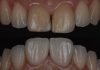
A patient presented to our clinic with a discomfort in the upper right central incisor (tooth 11) treated 20 years ago.
After clinical and radiographic examination (external resorption), the treatment plan involved extracting the tooth, immediately placing an implant, and using the extracted tooth’s crown as a temporary restoration. The situation was explained to the patient, and he agreed to the treatment plan.
The challenge in such cases is the successful restoration of tooth 11. To achieve optimal aesthetic results, we decided to do the “implant veneering approach” and place a veneer on the adjacent upper left central incisor (tooth 21).

Fig.2
On the same day as the implant procedure, the tooth was carefully extracted to avoid fracturing any part of the alveolar bone.
The external resorption was clearly visible between the cervical and middle parts of the root.

Fig.3
A lateral view of the extracted tooth shows the external resorption from another angulation.

Fig.4
After extracting the tooth and removing the inflammatory tissue, the socket was ready to be prepared for immediate implant placement.
Fig.5
The socket preparation was performed using MotorSurg (Eighteeth, Changzhou, China).
After the preparation, the implant was placed with a good primary stability.

Fig.6
View of the implant after immediate placement.
To prevent loss of the buccal bone and avoid anterior gum depression, the buccal socket preservation technique was employed.

Fig.7
To preserve the buccal bone of the socket, a fragment of the root is placed between the implant and the bone. This technique prevents anterior gum depression after bone remodeling.

Fig.8
A view of the implant, with the root fragment in place, to preserve the buccal socket.

Fig.9
The crown of the extracted tooth was used as a long-term provisional after placing the provisional abutment. This crown was bonded to the adjacent teeth with minimal occlusal load.

Fig.10
The provisional was bonded to the adjacent teeth on the palatal side using an orthodontic wire for retention.
The patient should be seen after three months.

Fig.11
The patient was seen after three months for evaluation and provisional preparation.
The same old crown was relined on the temporary abutment, placed slightly below the gum level, with a bulky cervical area. This approach guides the gum to migrate and reach the level of the provisional, establishing the future limit of the final crown.
The patient will be seen after four weeks.

Fig.12
After one month, the patient was seen, and the gum had reached the level of the provisional, as predicted. At this time, an impression was taken to prepare the zirconia crown at the implant level.

Fig.13
After one month, the gum had healed and migrated cervically, following the temporary profile.
At this step a digital impression was done using Helios 500 (Eighteeth, Changzhou, China) intra oral scan, to fabricate the crown over implant.

Fig.14
After a few days, a zirconia crown (Aidite, Qinhuangdao, China) was fabricated and tried in to ensure a proper fit. Note that the zirconia was prepared with the intention of bonding a veneer over it.
Restoring a single central incisor is quite challenging. To achieve a successful aesthetic result, the decision was made to place two lithium disilicate veneers on tooth 21 and the core of tooth 11.


Fig.15
From an occlusal view, we can see the hole in the crown for screwing it onto the implant. Note the space left for the buccal veneer. Tooth number 21 will be minimally prepared to create space for the veneer.

Fig.16
A buccal view showing the minimal preparation of tooth 21.

Fig.17
A palatal view showing the space left on teeth number 11 and 21.

Fig.18
An analog impression was taken with the zirconia crown in place, allowing the lab technician to prepare the veneers with precision.

Fig.19
A provisional abutment was used again on the implant to hold the provisional restoration, which was attached to the provisional on tooth number 21.

Fig.20
A lateral view of the crown shows the emergence profile of the crown over the implant, along with the preparation for the buccal veneer.

Fig.21
The slide shows the final crown over the implant, along with the final zirconia veneers. The Zirconia material used is EZneer (Aidite, Qinhuangdao, China) , treated with Biomic LiSi Connect (Aidite, Qinhuangdao, China) for optimal bonding. This LiSi layer, applied on the zirconia surface to bond, before sintering, enhances the clinical bonding performance of zirconia, providing an effect similar to glass ceramics.

Fig.22
After fixing the crown on the implant, the two disilicate veneers were bonded. The final result shows the similarity between the two central incisors.

Fig.23
A lateral view of the case shows the bulky restoration on the cervical part of the two veneers. This design protects the gum from food impaction and prevents future retraction.
Conclusions
In conclusion, the aesthetic restoration of a central incisor following extraction due to external resorption presents a significant clinical challenge. The combination of an immediate implant placement and the use of lithium disilicate veneers on adjacent teeth offers an effective solution. This approach not only ensures the stability and functionality of the implant but also achieves optimal aesthetic results by maintaining the harmony and symmetry of the smile. By carefully planning and executing the implant placement and veneer bonding, clinicians can restore both the appearance and confidence of the patient, demonstrating the transformative potential of modern dental techniques in addressing complex cases.
Bibliography
- Loi, E. (2013). Biologically oriented preparation technique (BOPT): a new approach for prosthetic restoration of periodontically healthy teeth. Eur J Esthet Dent, 8(1), 10-23.
- Ribeiro Martin, Leandro Lécio de Lima Sousa, Tiago Garcia Margu. “Aesthetic Rehabilitation with a Dental Implant in an Upper Central Incisor.” SVOA Dentistry, 2021.
- Kalghoum, I., Azzouzi, I., Hadyaoui Dalenda, B., Belhssan, H., & Cherif, M. (2023). “Guidelines for restoring fractured central incisors.” OAText, 10(1), 113-123.