Adhesive cementation is a critical step in the longevity of indirect posterior restorations. Successful outcomes depend on accurate diagnosis, proper tooth preparation, effective isolation and a simplified adhesive protocol.Contemporary universal adhesive systems and resin cements can help simplify the cementation workflow by reducing the number of clinical steps while maintaining a reliable adhesion. This clinical case illustrates a complete adhesive workflow for the replacement of a defective posterior restoration using Scotchbond™ Universal Plus Adhesive and RelyX™ Universal Resin Cement.


Fig.1
Preoperative radiographic assessment showing the extent of the carious lesion and supporting treatment planning.

Fig.2
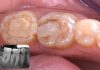
Initial clinical situation showing a defective posterior restoration with extensive structural compromise requiring adhesive rehabilitation.

Fig.3
The defective restoration and caries was carefully removed while preserving the maximum amount of healthy tooth structure.

Fig.4
Rubber dam isolation was established to provide optimal moisture control throughout the adhesive procedure.

Fig.5
Careful removal of residual restorative material and unsupported tissues while preserving sound dental structure.

Fig.6
Cavity configuration after selective tissue preservation and before final preparation for adhesive inidrect rehabilitation.

Fig.7
A matrix was placed deep into the sulcus to achieve isolation of the deep margin and facilitate anatomical reconstruction.

Fig.8
Selective enamel etching was performed to optimize adhesion while preserving the benefits of the universal adhesive strategy.

Fig.9
Application of Scotchbond™ Universal Plus Adhesive following the manufacturer’s recommended protocol under complete isolation.


Fig.10
Radiographic verification confirming complete removal of the previous restoration and adequate cavity preparation before the indirect restorative phase.

Fig.11
Digital evaluation of the restorative design, allowing verification of morphology, occlusion, and marginal integrity before fabrication of the overlay restoration.

Fig.12
Hydrofluoric acid etching of the internal ceramic surface to create a microretentive substrate for adhesive cementation.

Fig.13
Scotchbond™ Universal Plus Adhesive was applied to the conditioned ceramic surface, eliminating the need for a separate silane application when used in combination with RelyX™ Universal Resin Cement.

Fig.14
Prepared tooth after adhesive preparation, ready for rubber dam isolation.

Fig.15
Clinical try-in confirming the adaptation and morphology of the definitive restoration before cementation.

Fig.16
Complete isolation ensured optimal moisture control during the adhesive cementation procedure.

Fig.17
A clinical try-in was performed under complete isolation to verify marginal integrity, proximal contacts, and occlusal adaptation before cementation.

Fig.18
Cleaning and air abrasion of the preparation with AquaCare® improved substrate quality before the adhesive cementation procedure.

Fig.19
Adjacent teeth were protected to avoid inadvertent surface treatment and preserve the integrity of the surrounding enamel during adhesive cementation.

Fig.20
Selective phosphoric acid etching of the enamel optimized adhesion while maintaining the simplified universal adhesive protocol.

Fig.21
Active application of Scotchbond™ Universal Plus Adhesive enhanced substrate infiltration, promoting a reliable adhesive interface before cementation.

Fig.22
RelyX™ Universal Resin Cement was used to simplify the cementation workflow while maintaining a predictable adhesive protocol.


Fig.23
Careful removal of excess resin cement before final polymerization to facilitate marginal finishing.

Fig.24
Final restoration demonstrating harmonious integration with the surrounding dentition and accurate occlusal morphology.

Fig.25
Occlusal view showing natural anatomy, well-defined occlusal morphology, and functional integration of the indirect restoration.

Fig.26
Postoperative radiographic evaluation confirming complete seating, precise marginal adaptation, and the absence of residual excess cement.

Fig.27
Final occlusal view confirming accurate anatomy, stable proximal contacts, and functional integration of the definitive restoration.
Conclusions
Simplifying the adhesive cementation protocol without compromising fundamental biological and adhesive principles allows clinicians to achieve predictable, durable, and minimally invasive posterior indirect restorations.
Careful substrate management, meticulous isolation, and the use of contemporary universal adhesive systems contribute to an efficient clinical workflow while ensuring excellent marginal integrity, functional performance, and long-term restorative success.
Bibliography
- Fradeani M, Barducci G. Esthetic Rehabilitation in Fixed Prosthodontics. Volume 2: Prosthetic Treatment. Quintessence Publishing; 2008.
- Veneziani M. Adhesive restorations in the anterior dentition: biomimetic principles and minimally invasive preparations. Int J Esthet Dent. 2017.
- Magne P, Belser UC. Bonded Porcelain Restorations in the Anterior Dentition: A Biomimetic Approach. Quintessence Publishing; 2002.
- Magne P. Immediate dentin sealing: a fundamental procedure for indirect bonded restorations. J Esthet Restor Dent. 2005;17:144–154.
- Van Meerbeek B, De Munck J, Yoshida Y, et al. Buonocore Memorial Lecture. Adhesion to enamel and dentin: current status and future challenges. Oper Dent. 2003;28:215–235.
- Breschi L, Mazzoni A, Ruggeri A Jr, et al. Dental adhesion review: aging and stability of the bonded interface. Dent Mater. 2008;24:90–101.
- Ferrari M, Vichi A, Grandini S. Efficacy of different adhesive techniques on indirect ceramic restorations. Am J Dent. 2001.