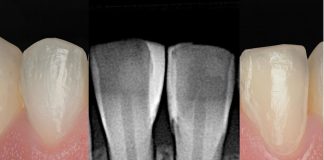
Direct pulp capping with calcium silicate-based materials has shown promising biological outcomes, especially when applied immediately after achieving hemostasis. A well-sealed permanent restoration significantly influences the success rate of vital pulp therapy. Indeed, when the final restoration is delayed to a second appointment, the risk of contamination and failure increases. Although Biodentine™ can be used as a temporary filling material, current evidence supports completing the restoration in a single session to minimize exposure to the oral environment.
To overcome the clinical limitation posed by the material’s setting time, this report introduces a new operative strategy that combines Biodentine XP with a self-adhesive restorative composite, allowing for immediate and definitive sealing in a single visit without compromising the biological activity of the calcium silicate-based materials. This protocol optimizes efficiency while preserving the biological benefits of pulp vitality conservation.


Fig.1
Initial situation, a 21-year-old patient presented with cold sensitivity in the upper right posterior region. Radiographic examination revealed multiple carious lesions of varying extent. In particular, the lesions on the first premolar and the first molar appeared deep and in close proximity to the pulp chamber. At the first appointment, the patient presented on an emergency basis due to pain. The two cavities were opened and temporarily sealed in order to relieve symptoms and assess pulp status. A second session was scheduled to allow for complete caries removal and definitive direct composite restorations. The periapical radiograph clearly shows the proximity of the lesions to the dental pulp for the first premolar. Indeed, a direct pulp capping procedure was considered, taking into account the patient’s young age to preserve the vitality of the tooth.

Fig.2
Starting with the first premolar, after removal of the temporary restoration and further cleaning of the cavity, a pulp horn was exposed. Once hemostasis was achieved, a direct pulp capping procedure was performed.

Fig.3
After proper application of a sectional matrix, Biodentine XP was applied in direct contact with the exposed pulp horn in a limited and controlled amount. Within the first 5 minutes, once adapted, it was covered using Vertise Flow (Kerr Dental), a self-adhesive flowable composite. This approach was intended to protect the material from subsequent adhesive procedures during definitive restoration, allowing it to set properly without compromising its biological and mechanical properties.

Fig.4
After polymerization of the Vertise Flow layer, the adhesive protocol was carried out. A universal adhesive containing the functional monomer 10-MDP (or its derivatives) was used. Selective enamel etching was performed for 20 seconds, followed by the application of the bonding agent.

Fig.5
Once the adhesive procedure was completed, the class II cavity was converted into a class I by rebuilding the proximal wall using a body-shade composite resin. To reproduce the correct height of the wall and avoid excessive occlusal material, the LM-Dental Misura Posterior instrument was used, assisting the clinician in optimizing anatomy and minimizing the need for occlusal adjustments.

Fig.6
The cavity was filled with a single increment of body-shade composite. Occlusal morphology was then finalized using the Espresso Posterior technique, enabling a fast and efficient completion of the direct restoration. Subsequently, the same treatment proceeded on the remaining two teeth.

Fig.7
Final situation immediately after rubber dam removal. All three teeth were definitively restored in a single session. A clinical and radiographic follow-up is essential to monitor the long-term outcome of the direct pulp capping on the first premolar, ideally up to at least 3 months after treatment.

Fig.8
The 3‑month radiograph revealed the absence of periapical lesions and confirmed the intact seal of the restorations, indicating an optimal radiographic outcome.

Fig.9
Clinical final aspect of three direct restoration after 3 months.
Conclusions
This clinical report presents an innovative strategy for managing direct pulp capping in a single session, overcoming the limitations associated with the setting time of tricalcium silicate-based materials. By integrating Biodentine XP with a self-adhesive restorative composite, it is now possible to complete both pulp protection and definitive restoration in a single clinical step. This protocol reduces the risk of microbial contamination linked to delayed restoration, offering a simplified, time-efficient, and biologically sound solution for vital pulp therapy.
Bibliography
- Celiksoz O, Irmak O. Delayed vs. immediate placement of restorative materials over Biodentine and RetroMTA: a micro-shear bond strength study. BMC Oral Health 24, 130 (2024).
- Camilleri J. Hydration characteristics of Biodentine and Theracal used as pulp capping materials. Dent Mater. 2014;30:709–15.
- Anastasiadis K, Koulaouzidou EA, Palaghias G, Eliades G (2018) Bonding of composite to base materials: effects of adhesive treatments on base surface properties and bond strength. J Adhes Dent 20:151–164.
- Falakaloğlu S, Yeniçeri Özata M, Plotino G. Microshear bond strength of different calcium silicate materials to bulk-fill composite. PeerJ. 2023 Mar 29;11:e15183. doi: 10.7717/peerj.15183.