The replacement of extensive amalgam restorations remains a common clinical challenge in restorative dentistry. Current adhesive concepts advocate a minimally invasive approach that preserves sound tooth structure while restoring biomechanical integrity through predictable adhesive protocols.
Advances in universal adhesives and contemporary resin composites have significantly expanded the indications for direct restorations, allowing clinicians to manage even severely compromised posterior teeth with excellent functional and aesthetic outcomes. This clinical report illustrates a conservative adhesive rehabilitation of a heavily restored posterior tooth, highlighting the importance of selective tissue preservation, immediate reinforcement of weakened cusps, and simplified composite layering to achieve long-term clinical success.


Fig.1
Sometimes, an XL restoration can unexpectedly turn into an XXL case during treatment.

Fig.2
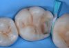
Initially, the case appeared relatively straightforward: a small defect in the buccal wall that seemed easy to restore with a flowable composite. A small amount of flowable material was used to repair the defect and smooth the wall.

Fig.3
After rubber dam isolation, a silicone index was fabricated to reproduce the original anatomy of the tooth and the existing amalgam restoration before its removal.

Fig.4
However, once the amalgam had been removed, it became evident that the defect was far more extensive than anticipated.

Fig.5
The adjacent tooth was also involved, making it ideal to restore it during the same procedure.

Fig.6
The palatal cusp lacked adequate enamel support and therefore required reduction. A visible crack line further confirmed the indication for cusp coverage.

Fig.7
At this stage, the previously fabricated silicone index could still be used. A try-in confirmed its accurate fit.

Fig.8
The silicone index was sectioned into two parts to separately reproduce the palatal and buccal cusps.

Fig.9
A conventional adhesive protocol was performed using Scotchbond™ Universal Plus Adhesive (Solventum).


Fig.10
The palatal cusp was restored first using the silicone index and Filtek™ One Bulk Fill Restorative composite (lateral view).


Fig.11
Occlusal view of the restored palatal cusp.

Fig.12
The buccal cusps were subsequently reconstructed using Filtek™ One Bulk Fill Restorative composite and the silicone index. At this stage, however, no space remained to properly shape the occlusal anatomy or reconstruct the interproximal walls.

Fig.13
To overcome this limitation, composite was selectively removed to create sufficient space for proper anatomical reconstruction.

Fig.14
The cavity was reopened, and the adhesive layer was reactivated.

Fig.15
The interproximal walls were reconstructed using the Polydentia GO ring and the Polydentia Flex matrix system.

Fig.16
At this stage, the case had effectively been simplified into a Class I restoration, transforming a complex reconstruction into a far more manageable procedure.

Fig.17
At this stage, the case had effectively been simplified into a Class I restoration, transforming a complex reconstruction into a far more manageable procedure.

Fig.18
Final restoration after finishing and polishing.

Fig.19
Final restoration with 6-month follow-up.
Conclusions
This clinical case demonstrates that even extensively restored posterior teeth can be predictably rehabilitated using a conservative adhesive approach.
Careful diagnosis, preservation of healthy tooth structure, immediate cusp reinforcement, and the use of contemporary adhesive materials allow clinicians to restore function, biomechanics, and aesthetics while delaying or avoiding more invasive indirect restorations. Adhesive dentistry continues to shift the restorative paradigm from replacement to preservation, reinforcing the philosophy that the best restoration is the one that conserves as much natural tooth structure as possible.


Bibliography
- Devoto W, Saracinelli M, Manauta J. Stratification techniques for direct posterior composite restorations. StyleItaliano Publications.
- Manauta J, Salat A, Putignano A, Devoto W, Bucciarelli A. Layers: An Atlas of Composite Resin Stratification. Quintessence Publishing.
- Putignano A, Devoto W, Manauta J. Direct posterior composite restorations: simplified approaches for predictable anatomy and aesthetics. International Dentistry.
- Spreafico R, Dietschi D. Restorative Treatment of the Posterior Dentition with Adhesive Techniques. Quintessence Publishing.
- Van Meerbeek B, Yoshihara K, Yoshida Y, Mine A, De Munck J, Van Landuyt KL. State of the art of self-etch and universal adhesives. Dental Materials. 2020.