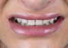
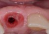
Following a tooth extraction, the alveolus do not always heal completely, reforming the entire alveolar process and thus re-establishing an ideal anatomy. Sometimes, also as a consequence of the severity of the cause for which the tooth was lost, they heal only partially, leaving defects that will need to be regenerated later to obtain an optimal result. In this clinical case a delayed post extractive implant was placed with contemporary bone regeneration to correct the vestibular bone dehiscence and to recreate the root bump (Root Bump Regeneration) in the first upper right premolar, in order to get a correct amount of bone around the implant, and a pleasant aesthetic result.


Fig.1
The initial X-ray shows a good amount of bone in the vertical direction, but don’t give any info about the bone width.

Fig.2
At the CBCT examination it is clear that there is a horizontal bone defect that needs to be treated with a bone regenerative surgery, and that the remaining bone is sufficient to insert an implant with a good probability to reach an ideal primary stability.

Fig.3
As the flap was raised, the bone defect was immediately evident.

Fig.4
After its insertion the implant showed a bone dehiscence in the buccal side. The defect was considered easy to treat because around the defect there was a good amount of bone.

Fig.5
To maintain a stable space around the defect it was decided to use a titanium mesh fixed directly to the implant by the surgical screw. The mesh was easily folded in order to recreate an anatomical ideal bone crest.
Then a series of holes were made to perforate the bone cortex and allow for greater bleeding to irrigate the graft and increase the supply of growth factors necessary for optimal healing.

Fig.6
The space between the mesh and the implant was filled with only heterologous biomaterial because the space to fill was little, and it was decided that the biomaterial alone would have been sufficient in this situation.

Fig.7
The mesh and the bone were then protected with a resorbable collagen membrane.

Fig.8
At the end of the surgery the flap was repositioned and sutured without any tension, and to facilitate the wound healing it was executed a flap without any vertical releasing incision.

Fig.9
X-Ray at the end of the implant placement.

Fig.10
The perfect healing at six months from the surgery. It is possible to see the mesh in transparency through the soft tissue.

Fig.11
The mesh was removed at six months from the first surgery, and at the reopening the mesh was clearly well integrated.

Fig.12
As soon as the grid was removed, excellent healing was evident, with the presence of a perfect bony balcony covered by a thick layer of pseudo-periosteal tissue.

Fig.13
The flap was then repositioned against the healing screw and slightly more apically to allow for the formation of a greater amount of keratinized gingiva.

Fig.14
One month after reopening, a digital impression was taken to convey to the technician the position of the implant and the prosthetic preparation of the adjacent natural tooth.

Fig.15
Ten years later the tooth looks absolutely natural, and the soft tissue surrounding it is absolutely healthy and pleasant.

Fig.16
The X-ray at 10 years shows a stable and well defined bone contouring the implant without any bone radio-transparency, confirming the success of the therapy.
Conclusions
The possibility to insert an implant in contemporary with the GBR procedures gives not only the possibility to reduce the surgical sections, but in contemporary it gives the possibility to create the root bump prominence, together with a correct soft tissue management and so, to reach a better and absolutely natural and esthetic result.
The use of a titanium mesh made it possible to ideally shape the regeneration volume, in order to optimise almost exactly the bone balcony, matching the adjacent bone position, and thus imitating as much as possible the original position of the bone tissue. In this way it is possible to get an aesthetic pleasant result, and the hard and soft tissues stability over time.
Bibliography
- Chu SJ., et al. “Subclassification and Clinical Management of Extraction Sockets with Labial Dentoalveolar Dehiscence Defects”. Compendium of Continuing Education in Dentistry 36.7 (2015): 516, 518-520.
- Carvalho W, Ladeira Casado P, Caúla AL, Porto Barboza E: Implants for single first molar replacement: important treatment concerns. IMPLANT DENT 2004;13:328-335.
- Proussaefs P, Lozada J. Use of titanium mesh for staged localized alveolar ridge augmentation: clinical and histologic-histomorphometric evaluation. J Oral Implantol. 2006;32(5):237-47. doi: 10.1563/1548-1336(2006)32[237:UOTMFS]2.0.CO;2. PMID: 17069168.