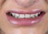
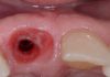
The first permanent molar is a tooth of primary importance for maintaining the shape of the arch and the correct occlusal system. It is the first tooth to erupt and, unfortunately, is often also the first to be extracted. The loss of this tooth almost always leads to an occlusal imbalance, which manifests itself with the mesialization and distalization of the adjacent teeth and the extrusion of antagonists. In the past, the only treatment options to replace the missing tooth involved the application of a removable prosthesis or a fixed bridge, with the compromising, in the latter case, of two adjacent, often healthy, teeth.
Nowadays, these two options have been joined by the possibility of inserting an endosseous implant that could support a crown and therefore restore chewing and esthetic.
In this clinical case an immediate post extractive implant placement with immediate provisionalization in the first lower left molar is combined with a contemporary regenerative procedures to fill the gap between the fixture and the surrounding bone.


Fig.1
The patient presented to our clinic complaining of an ill-defined pain during chewing on tooth 36.
The X-ray show a radiolucency apically to the mesial root, and at the clinical examination a point probing was found at the vestibular – mesial root site, revealing the presence of a root fracture.
The crown and the soft tissue of the tooth are still pleasant for shape, contour and tone. So, they are a base to start, to respect and to maintain during all the phases of the treatment, and the well
represented interproximal bone crest with the large inter-radicular septum are decisive to take the decision to extract the tooth and to immediate insert an implant.

Fig.2
The patient presented to our clinic complaining of an ill-defined pain during chewing on tooth 36.
The X-ray show a radiolucency apically to the mesial root, and at the clinical examination a point probing was found at the vestibular – mesial root site, revealing the presence of a root fracture.
The crown and the soft tissue of the tooth are still pleasant for shape, contour and tone. So, they are a base to start, to respect and to maintain during all the phases of the treatment, and the well represented interproximal bone crest with the large inter-radicular septum are decisive to take the decision to extract the tooth and to immediate insert an implant.

Fig.3
After having removed the ceramic crown, the tooth has been separated in two portions along its furcation to facilitate the extraction.

Fig.4
Immediately after the extraction it is evident the almost favorable anatomy of the septum.

Fig.5
The implant has been positioned into the septum and slightly lingual, so to maintain 2-3mm between it and the buccal bone vestibular plate. It is visible that the mesial and distal portions of the implant are out of the septum bone (type B socket).

Fig.6
After the provisional relining, the residual socket has been filled with some bone substitute up to the gingival margin, with the objective to reduce the bone remodeling and to sustain the soft tissues.

Fig.7
The screwed provisional crown has a concave shape, in its submerged portion, starting exactly at the finishing line of the old provisional crown.

Fig.8
The provisional crown just screwed at the end of the surgery. Its concave shape totally respects the soft tissues without giving them any compression or ischemia sign. Its perfect contour allows to maintain the soft tissues exactly in the same position in which they were before the surgery. A single suture has been given because the gingival margin was lacerated during the extraction operation.

Fig.9
6 months after the surgery, the tissue are still in their original position, the papillae and the parabola are filling correctly their space around the provisional restoration, and the root bump is well represented as it was before the molar extraction, giving to the soft tissue surrounding the restoration a perfect natural aspect.

Fig.10
As the screwed provisional crown has been removed in order to take the impression, the tissues show their perfect anatomic shape and contour preservation, and some biomaterial particles are still visible inside the soft tissue without any inflammation sign.

Fig.11
According to Hinds, a custom made transfer, exact replica of the trans-mucosal portion of the provisional crown, is used to take an impression.

Fig.12
6 months after definitive crown screwing (1 year from the surgery) it is evident the quality, the shape and the tone of the soft tissues.

Fig.13
They have almost maintained their original shape and contour. The root bump is well preserved avoiding food entrapment and greyish discoloration.

Fig.14
The final X-ray shows the bone stability along with the bone substitute reorganization.
Conclusions
Dilemmas accompanying implant in the molar area frequently involve the choice if would it better to maintain or not the root bump anatomy.
The article presents a case of immediate post extractive implant placement with immediate provisionalization in the first lower left molar in contemporary with a regenerative procedures to fill the gap between the fixture and the surrounding bone with heterologous bone substitute.
Nowadays, a single implant supported restoration has to answer and provide aesthetic and function especially when a single element has to be replaced in the anterior sector, but also in the posteriors. The therapeutic choice adopted has led the clinician to an ideal management of the original anatomy in term of hard and soft tissue, facing against the typical bone contraction when the tooth is not in site. It was possible to observe and figure out at the end of the process that the preservation of the natural root bump could be useful also for the cleansing and maintenance of the rehabilitation, because it prevents the typical food entrapment between the crown and the gingival profile, besides the aesthetic and the pleasant result. It should be interesting to investigate if this procedure is needed only for esthetic reasons or if the soft tissue’s anatomy shape preservation could facilitate the daily maintenance for the patients, improving the long lasting of the implant and the prosthetic rehabilitation as we believe.
Bibliography
- Carvalho W, Ladeira Casado P, Caúla AL, Porto Barboza E: Implants for single first molar replacement: important treatment concerns. IMPLANT DENT 2004;13:328-335.
- Saito H, Chu S, Reynolds M, Tarnow D. Provisional Restorations Used in Immediate Implant Placement Provide a Platform to Promote Peri-implant Soft Tissue Healing: A Pilot Study. Int J Period Rest Dent 2016;36:47-52.3.
- Smith RB, Tarnow DP. Classification of molar extraction sites for immediate dental implant placement: technical note. Int J Oral Maxillofac Implants 2013;28: 911-6.