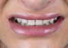
Whenever possible, our first treatment option is immediate post-extraction implant placement with immediate provisionalization. Sometimes, for a variety of reasons, this option is not feasible, so we may decide to extract the fractured tooth, and in a second time, after the infection is healed, to proceed with the implant placement together with the bone regeneration and the contemporary provisionalization.
In this clinical case a delayed post extractive implant was placed with immediate provisionalization and contemporary bone regeneration to correct the vestibular bone fenestration and to recreate the root bump in the first upper right premolar, in order to get a correct amount of bone around the implant, and a pleasant aesthetic result.


Fig.1
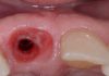
Three months after the first premolar extraction, the site is healed and ready for the surgery. It is clearly evident a horizontal minus in the buccal side, expression of a probably bone contraction.

Fig.2
The X-Ray at three months after the tooth extraction, it shows an incomplete bone healing.

Fig.3
After the implant placement there was a bone dehiscence in the buccal side. The defect was favourable because it was a four wall defect. One of the most easy to treat to get a complete buccal bone regeneration.

Fig.4
Before to go with the GBR a provisional crown was relined and screwed, then a series of holes were made to perforate the bone cortex and allow for greater bleeding to irrigate the graft and increase the supply of growth factors necessary for optimal healing.

Fig.5
The defect was filled with heterologous biomaterial.

Fig.6
And was then protected with a resorbable collagen membrane.

Fig.7
At the end of the surgery the flap was repositioned and sutured without any tension against the provisional restoration.

Fig.8
X-Ray at the end of the implant placement.

Fig.9
At six months from the surgery an impression according with the Hinds method was taken to accurately replicate the transmucosal path created with the provisional restoration.
This is an old case, now a digital impression with the same intention is used.

Fig.10
3 years later the first premolar crown is still pleasant. Its emergence profile looks natural, and it is indistinguishable from that of the second premolar. The root prominence remained stable, creating a completely natural effect.

Fig.11
The X-Ray at 16 years.

Fig.12
CBCT at 3 years shows a stable and well defined buccal bone plate, confirming the success of the therapy.

Fig.13
CBCT at 16 years shows a stable and well defined buccal bone plate, confirming furthermore the success of the therapy.
Conclusions
The possibility to insert an implant in contemporary with the GBR procedures and its immediate provisionalization gives not only the possibility to reduce the surgical sections, but in contemporary it gives the possibility to create the root bump prominence, together with a correct soft tissue management and so, to reach a better and absolutely natural and esthetic result.
Bibliography
- Chu SJ., et al. “Subclassification and Clinical Management of Extraction Sockets with Labial Dentoalveolar Dehiscence Defects”. Compendium of Continuing Education in Dentistry 36.7 (2015): 516, 518-520.
- Hinds KF. “Custom impression coping for an exact registration of the healed tissue in the esthetic implant restoration”. International Journal of Periodontics and Restorative Dentistry 17.6 (1997): 584-591.
- Araujo MG., et al. “Bio-Oss collagen in the bucal gap at immediate implants: a 6 months study in the dog”. Clinical Oral Implants Research 22.1 (2011): 1-8.