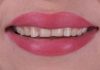
A clinical case by our Community member Dr. Eyup Celik
This article and its content are published under the Author’s responsibility as an expression of the Author’s own ideas and practice. Styleitaliano denies any responsibility about the visual and written content of this work.
Restoring a harmonious smile and achieving optimal functional occlusion are essential objectives in modern dentistry. Advances in adhesive dentistry and ceramic materials, particularly lithium disilicate veneers, have made it possible to perform comprehensive aesthetic and functional rehabilitations with minimally invasive techniques.
This case report presents the treatment of a patient with compromised dental aesthetics and reduced vertical dimension of occlusion (VDO) using 28 units of lithium disilicate veneers. The patient’s chief complaint included dissatisfaction with her smile and functional discomfort associated with occlusal wear. The approach involved increasing the vertical dimension of occlusion to restore facial balance, improve functionality, and enhance the smile’s aesthetic appeal.
The following report details the diagnostic process, treatment planning, and clinical execution, highlighting the advantages of lithium disilicate veneers in achieving a predictable and durable outcome.


Fig.1
A 45-year-old female patient presented to our clinic with the complaint that her teeth were not visible when speaking and that she was dissatisfied with her smile. In the first photograph, taken with the lips in a resting position, the upper teeth were completely hidden, contributing to an aged appearance. The second photograph was taken while the patient was smiling.

Fig.2
Additional photographs were taken to analyze tooth display from a cross-sagittal view.
Video.1
In such cases, video recordings can enhance the collaboration between the clinician and the dental technician by aiding in the analysis of the smile, the harmony between the lips and teeth, and the determination of the ideal position and extent of the new smile design.

Fig.3
Intraoral photographs were taken to assess the occlusal plane and evaluate the intraoral condition. Clinical examination revealed existing porcelain onlays on the premolars and molars of the lower jaw. Additionally, an implant-supported crown was present on tooth 36.

Fig.4
Long-term bruxism resulted in generalized tooth wear, a reduced vertical dimension, and dental misalignment. Clinical findings included widespread abrasion and flattening of the occlusal and incisal surfaces, with areas of exposed dentin. Mild misalignment was observed in the upper lateral and central incisors.

Fig.5
Occlusal view of the lower arch.

Fig.6
A functional and motivational mock-up was applied a few days after the initial appointment, during which photographs and impressions were taken. The purpose of the mock-up was to evaluate the proposed smile design in line with the patient’s aesthetic expectations, to confirm patient satisfaction, and to functionally assess the new bite registration.

Fig.7
Close-up photographs were taken of the mock-up while the patient was giving a full smile.



Fig.8
Intra-oral bite registration.
Video.2
A video recording was made to ensure that the mock-up application was fully understood by the technician and to provide accurate guidance for reproducing the correct tooth size and volume.

Fig.9
Tooth preparation was performed carefully under a microscope using a minimally invasive approach. Following preparation, retraction cords were placed into the sulcus, and polyether impressions (3M Impregum by Solventum) were taken for both the upper and lower teeth. New bite records were also obtained. Bis-acryl temporary restorations were then applied, which the patient would wear until the bonding phase of the lithium disilicate veneers (E-max, Ivoclar Vivadent) at her next appointment.

Fig.10
After bonding the final restorations, full-face photographs of the patient smiling were taken from various angles. The treatment not only enhanced the aesthetics of her smile but also significantly improved her self-confidence.

Fig.11
The final result after bonding the veneers showed a successful aesthetic and functional outcome. In the premolar and molar regions, the veneers were fabricated as monolithic lithium disilicate restorations, while the anterior veneers were created using the layering technique with pressed lithium disilicate (E-max) for enhanced translucency and natural appearance. The vertical dimension of occlusion was increased by 5 mm in the anterior region.

Fig.12
Close-up photographs of the new smile show the harmony between the veneers and the patient’s lips.
Video.3
Following the bonding of the new veneers, dentofacial harmony was reestablished, and proper tooth visibility was restored. As a result, the patient regained a more youthful and vibrant appearance.

Fig.13
Occlusal views of the table-top veneers demonstrate the approach used to increase the vertical dimension. The vertical dimension was restored by restoring both the upper and lower teeth.
Conclusions
This case highlights the transformative potential of minimal-preparation veneers in addressing worn dentition, arch discrepancies, and aesthetic concerns—while preserving natural tooth structure. By combining functional rehabilitation with aesthetic precision, optimal long-term outcomes can be achieved.
Restoring the vertical dimension of occlusion (VDO) plays a crucial role in dentofacial harmonization. Increasing the VDO contributes to a more balanced facial profile, reducing the collapsed appearance often associated with extensive tooth wear. This not only rejuvenates the patient’s overall appearance but also improves functional aspects such as occlusal stability and masticatory efficiency. Properly addressing VDO through minimally invasive techniques, like veneers, ensures long-term success while preserving natural tooth structure.
Bibliography
- Koubi S, Gurel G, Margossian P, Massihi R, Tassery H. A Simplified Approach for Restoration of Worn Dentition Using the Full Mock-up Concept: Clinical Case Reports. Int J Periodontics Restorative Dent. 2018 Mar/Apr;38(2):189-197.
- Vailati F, Belser U: full month adhesive rehabilitation of a severely eroded dentition: 3 step technique part 2 Eur J Esthet Dent. 2008 Summer;3(2):128-46.