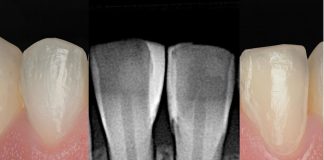
A 30-year-old female patient presented to our clinic complaining of an evident esthetic issue involving tooth 22 (maxillary left lateral incisor). The patient’s main concern was the chromatic discrepancy of this tooth compared to the adjacent dentition, which negatively affected her smile aesthetics.
Clinical examination revealed that tooth 22 was endodontically treated and appeared darker than the surrounding teeth.

Fig.1
Additionally, the buccal enamel exhibited increased roughness and a central dark discoloration consistent with localized demineralization. No functional problems were detected, and the periodontal status was within normal limits.
Given the predominantly esthetic nature of the case, a minimally invasive treatment plan was proposed.
The plan included professional dental prophylaxis, followed by at-home whitening of the entire maxillary arch, with increased focus on tooth 22 and the canines. After the whitening phase, a direct esthetic composite restoration of tooth 22 was planned in order to restore correct morphology and improve color integration with the adjacent teeth.

Fig.2
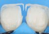
The first clinical step consisted of shade selection using a reference shade tab.


Fig.3
After approximately 2–3 weeks of at-home whitening, performed using Whiteness Perfect 16% carbamide peroxide (FGM) according to a dedicated bleaching protocol for the entire maxillary arch and enhanced application on selected teeth, a significant improvement in overall tooth color was observed.

Fig.4
The chromatic discrepancy of tooth 22 was notably reduced, creating a favorable substrate for the subsequent restorative phase.

Fig.5
The operative field was then isolated using a rubber dam, with particular attention to tooth 22.

Fig.6
A B4 rubber dam clamp was used to achieve precise isolation of the treated tooth, ensuring optimal moisture control.
A minimal preparation at the midline was performed using a diamond round bur, limited exclusively to the discolored and demineralized area, in accordance with a conservative approach.

Fig.7
Enamel etching was carried out using phosphoric acid, followed by thorough rinsing and air drying.

Fig.8
A universal adhesive system was applied using a microbrush, ensuring even coverage of the etched enamel surface, and subsequently light-cured according to the manufacturer’s instructions.

Fig.9
In this case the Ambar Universal APS adhesive from FGM was used.

Fig.10
For the restorative phase, a single shade of Vittra APS Unique composite resin from FGM was selected.
Thanks to its pronounced chameleon effect, this material allows optimal color blending with the surrounding tooth structure, making it particularly suitable for anterior esthetic restorations.

Fig.11
The composite was applied using the LM Solo Anterior spatula, which enables precise modeling and controlled pressure during placement, minimizing the risk of air bubble incorporation.

Fig.12
After polymerization, the restoration was evaluated for anatomical accuracy.

Fig.13
Finishing and polishing were performed using dedicated finishing burs and discs to refine the buccal morphology and recreate correct angle lines.

Fig.14
During the correction of the buccal surface morphology, the presence of composite dust provided valuable visual feedback regarding surface anatomy and transition line symmetry.
Low-speed instrumentation without water spray proved to be an effective technique for fine morphological adjustments.

Fig.15
Final evaluation included a direct comparison between pre-operative and post-operative conditions, showing a harmonious and natural esthetic result.
Conclusions
This clinical case highlights the effectiveness of combining tooth whitening with direct composite restoration in managing esthetic challenges involving discolored anterior teeth. Composite materials such as Vittra APS Unique from FGM, characterized by its chameleon effect, represent an ideal solution when restoring aesthetic areas, where seamless integration with the underlying tooth color is essential.
In this case, the whitening phase—performed using Whiteness Perfect 16% carbamide peroxide (FGM)—played a crucial role in achieving an optimal base color for tooth 22 and 23, allowing the restorative material to maintain and enhance the achieved brightness rather than masking an unfavorable substrate. The result demonstrated excellent chromatic integration, ease of handling, and efficient polishing characteristics.
What may initially appear as a complex esthetic case—especially in patients with high esthetic expectations and a tooth clearly out of chromatic harmony—can be predictably managed through a structured and minimally invasive approach. Incorporating a whitening phase prior to restorative treatment consistently aids clinicians in achieving superior, natural, and predictable esthetic outcomes.
Bibliography
- Orilisi G, Tosco V, Monterubbianesi R, Notarstefano V, Özcan M, Putignano A, Orsini G. ATR-FTIR, EDS and SEM evaluations of enamel structure after treatment with hydrogen peroxide bleaching agents loaded with nano-hydroxyapatite particles. PeerJ. 2021 Jan 29;9:e10606.
- Manauta J, Salat A. Layers, An atlas of composite resin stratification. Quintessence Publishing Co; 2012.
- Goodacre CJ, Eugene Roberts W, Munoz CA. Noncarious cervical lesions: Morphology and progression, prevalence, etiology, pathophysiology, and clinical guidelines for restoration. J Prosthodont. 2023;32:e1–e18. https://doi.org/10.1111/jopr.13585.
- Akal N, Over H, Olmez A, Bodur H. Effects of carbamide peroxide containing bleaching agents on the morphology and subsurface hardness of enamel. J Clin Pediatr Dent. 2001;25(4):293-6.
- Manauta J. The Closing Gap Technique. The perfect seal in Class 5 restorations. StyleItaliano
- Zanolla J, Marques A, da Costa DC, de Souza AS, Coutinho M. Influence of tooth bleaching on dental enamel microhardness: a systematic review and meta-analysis. Aust Dent J. 2017;62(3):276-282.