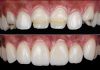
A clinical case by our Community member Dr. Rim Bourgi
This article and its content are published under the Author’s responsibility as an expression of the Author’s own ideas and practice. Styleitaliano denies any responsibility about the visual and written content of this work.
Tooth whitening remains one of the most sought-after esthetic procedures in contemporary dentistry, valued for its non-invasive nature, efficiency, and predictability. A range of techniques is available to meet varying patient needs, including in-office bleaching, dentist-supervised at-home treatments, and over-the-counter products. Home whitening systems, in particular, have gained popularity due to their convenience and cost-effectiveness, while still delivering professional-level results when properly prescribed.
This article presents a step-by-step clinical case that illustrates the indications, application protocol, and esthetic outcomes of a dental bleaching procedure. High-quality dental photography was employed to document and reliably analyze the evolution of the treatment, enhancing communication, clinical evaluation, and providing a foundation for further research on objective monitoring of dental procedures.

Fig.1
A 32-year-old female patient presented with a request for a non-invasive solution to address tooth discoloration. As part of the initial clinical evaluation, an intraoral photograph was taken with the patient in occlusion using retractors, providing a clear baseline image of the dental condition. This intraoral photograph was captured with the Smile Lite MDP2 (Smile Line, St-Imier, Switzerland), to enhance diagnostic accuracy and serve as a visual reference for treatment progression. Such photographic documentation is a fundamental component of modern dental practice, facilitating diagnosis, treatment planning, and outcome assessment.
Composite attachments were clearly visible on the upper posterior teeth, as the patient had previously undergone orthodontic treatment. Since she was planning to get married, the esthetic treatment plan was initiated.
The first phase of treatment consisted of a professional dental prophylaxis to remove extrinsic stains, plaque, and debris from the enamel surface. This step is essential in optimizing the effectiveness of whitening procedures and improving overall oral health.

Fig.2
One week later, the patient returned seeking a brighter smile. A bleaching procedure was performed, and a baseline frontal photograph was captured to document the initial dental condition. The image was taken using the Smile Lite MDP2 (Smile Line, St-Imier, Switzerland) at full power with a Samsung smartphone. To ensure soft, evenly diffused lighting, all six LEDs on each lateral panel and the eight LEDs on the central panel were activated with the white diffuser in place.

Fig.3
Another essential image can be captured by activating all light sources without any obstructions. This setup ensures uniform illumination from all angles, producing a glossy effect that enhances surface detail and clarity without requiring further brightness adjustments.

Fig.4
The initial tooth shade was assessed and verified using the VITA Classical Shade Guide, confirming a baseline shade of A2 prior to the commencement of the whitening procedure.

Fig.5
Image Captured with Polarizing Filter: This photographic technique employs a polarizing filter to eliminate surface reflections, resulting in an image that provides precise visualization of tooth color, brightness, and subtle surface details. Polarized photography is particularly valuable for accurately comparing tooth shade to the reference guide and for enhancing diagnostic clarity when assessing discoloration or planning esthetic treatments.

Fig.6
Artistic photograph depicting the initial dental situation.

Fig.7
Custom-fit trays can be fabricated either conventionally or digitally using an intraoral scanner, such as the Helios 500 (Eighteeth, Changzhou, China). In this case, a digital workflow was employed: a digital impression was captured, the tray was designed using exocad software (Exocad GmbH, Darmstadt, Germany), and then three-dimensional (3D)-printed for use.

Fig.8
The intraoral photograph, taken with the teeth in occlusion, clearly illustrates the level of whiteness achieved. It offers an accurate visual representation of the final shade obtained by the patient.

Fig.9
The final frontal view provides a comprehensive visual record of the patient’s whitening outcome.

Fig.10
After two weeks of treatment, the patient expressed satisfaction with the outcome, as clearly demonstrated in the full-light intraoral photograph.

Fig.11
Following two weeks of bleaching, the final outcome revealed a significant shade transformation—from an initial A2 to B1—closely matching the natural tooth color. This improvement was documented through an intraoral photograph, highlighting a highly satisfactory and esthetically pleasing result.

Fig.12
Later view of the final situation.
Several studies have evaluated the effectiveness of whitening systems on composite materials, but the analysis was generally limited to the material surface. While whitening procedures are effective in reducing surface pigmentation, the degree of color change depends on the staining solution and the material’s composition. Some authors have suggested that the observed color change is due to a superficial cleaning effect rather than an intrinsic whitening of the composite. However, the results of the present case do not support this view, as the whitening effect was also observed in the subsurface layers of the composite.
The mechanism of tooth whitening relies on hydrogen peroxide molecules, released from bleaching gels, penetrating dental tissues and interacting with the organic compounds responsible for pigmentation (chromophores). The interprismatic spaces in enamel and the dentinal tubules in dentin allow fluid movement, making dental hard tissues highly permeable and enabling hydrogen peroxide to exert its effect.
Although less is known about how bleaching agents interact with the 3D polymer network of dental composites, Durner et al. proposed that whitening products may cause oxidative cleavage of this network. This process could facilitate the diffusion of bleaching molecules into the composite structure, providing a plausible explanation for the subsurface whitening observed in this case.

Fig.13
The contrast between the “before” and “after” stages of dental bleaching is striking, enhancing not only the esthetic appearance of the teeth but also significantly boosting the patient’s confidence.

Fig.14
The intraoral photographs with retraction highlight the clear difference between the pre- and post-whitening stages, showcasing the improved dental esthetics and reflecting the patient’s increased satisfaction with her smile.

Fig.15
An extraoral photograph was captured using mobile dental photography to accurately document the post-whitening results. This approach not only provides a reliable visual record for clinical evaluation but also enhances patient communication. The patient expressed high satisfaction with the outcome and inquired about strategies to maintain the achieved level of whiteness over time.

Fig.16
The brilliance of the whitened teeth is clearly evident, a result of using the appropriate product combined with an effective technique.
Optimal use of the Smile Lite MDP2 (Smile Line, St-Imier, Switzerland) enabled precise control of lighting, resulting in high-quality, visually striking images. In this case, the patient was so satisfied with the outcome that she requested the photographs to share with her friends.

Fig.17
Frontal view of the teeth before and one month after the bleaching procedure, demonstrating the achieved shade improvement.

Fig.18
When performing mobile dental photography with auxiliary lighting, it is essential to follow standardized protocols to ensure reproducibility, accuracy, and high-quality images, as described by Prof. Louis Hardan (Protocols for Mobile Dental Photography with Auxiliary Lighting). In this case, extraoral (eye-level) photographs were captured to document the patient’s eyes. Proper positioning, consistent lighting using devices such as the Smile Lite MDP2, and careful attention to framing are key to achieving clinically useful and visually appealing images.
Conclusions
This clinical case highlights the integration of mobile dental photography within a digital dentistry workflow for tooth whitening assessment. High-quality extraoral and intraoral photographs provide a reliable baseline and enable precise, objective monitoring of shade changes over time. When combined with digital tools such as intraoral scanners, 3D software, and auxiliary lighting devices, mobile photography enhances reproducibility, accuracy, and documentation of clinical outcomes. Beyond improving patient communication and engagement, this approach demonstrates how digital technologies can elevate esthetic dentistry by enabling precise treatment planning, objective evaluation, and long-term follow-up.
Bibliography
- Durner J, Stojanovic M, Urcan E, Spahl W, Haertel U, Hickel R, Reichl FX. Effect of hydrogen peroxide on the three-dimensional polymer network in composites. dental materials. 2011 Jun 1;27(6):573-80.
- Giachetti L, Russo DS, Nieri M, Cinelli F. Can discolored dental composites be bleached in depth?. Restorative Dentistry & Endodontics. 2024 Jun 11;49(3):e(23).
- Salehi A, He M, Hampé-Kautz V, Etienne O. Digital evaluation of dental bleaching using a new methodology: an in vivo study. International Journal of Esthetic Dentistry. 2022 Dec 1;17(4):448-467.
- Hardan L, Bourgi R, Cuevas-Suárez CE, Lukomska-Szymanska M, Monjarás-Ávila AJ, Zarow M, Jakubowicz N, Jorquera G, Ashi T, Mancino D, Kharouf N. Novel trends in dental color match using different shade selection methods: a systematic review and meta-analysis. Materials. 2022 Jan 8;15(2):468.
- Hardan L. Protocols for mobile dental photography with auxiliary lighting. 2020.
- Devoto E, Devoto W, Pradella S, Enrique Cuevas-Suárez C, Eliezer Zamarripa-Calderon J, Dimitriu B, Lukomska-Szymańska M, Kharouf N, Bourgi R, Hardan L. Maintenance of Dental Bleaching: A Systematic Review. International Arab Journal of Dentistry. 2024 Jul 1;15(2).
- https://www.styleitaliano.org/document-bleach-maintain-dbm-a-clinical-protocol-for-managing-localized-black-stains-and-enhancing-whitening-results/
- https://www.styleitaliano.org/mobile-dental-photography-part-i/
- https://www.styleitaliano.org/three-magical-tools-for-successful-dental-bleaching/